. 2023 Jan;299(1):102797.
doi: 10.1016/j.jbc.2022.102797. Epub 2022 Dec 14.

The N-terminal domain of human mitochondrial helicase Twinkle has DNA-binding activity crucial for supporting processive DNA synthesis by polymerase γ

Affiliations

The N-terminal domain of human mitochondrial helicase Twinkle has DNA-binding activity crucial for supporting processive DNA synthesis by polymerase γ

Laura C Johnson et al. J Biol Chem. 2023 Jan.

Abstract

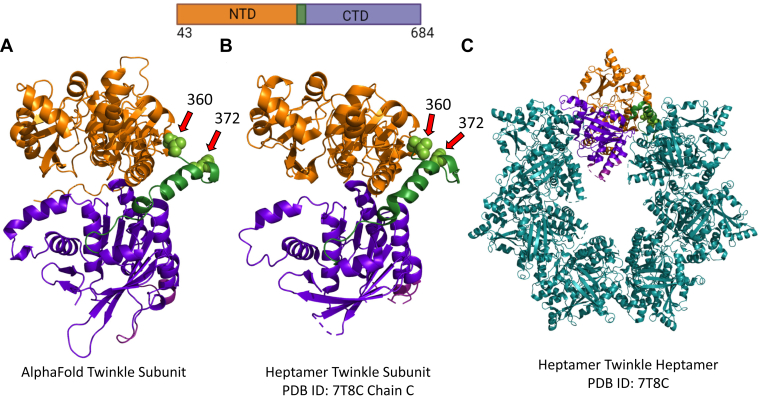
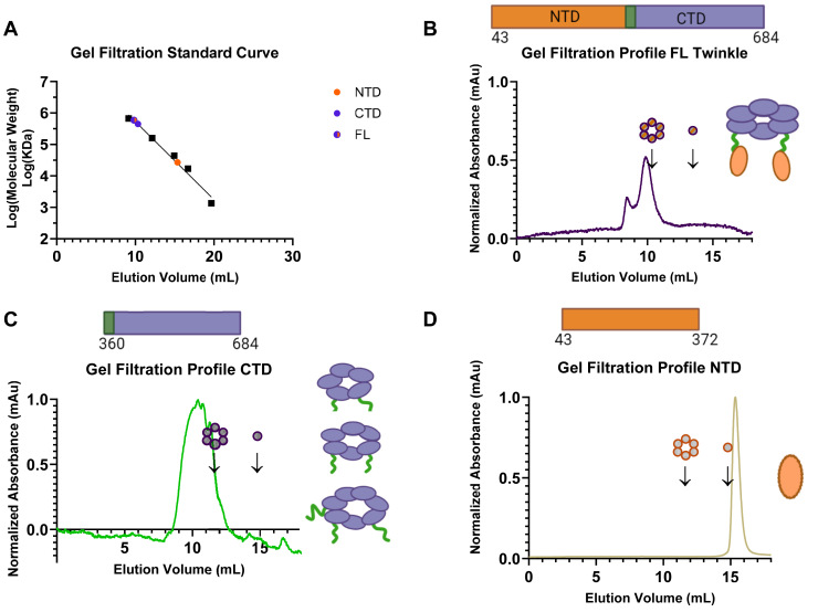
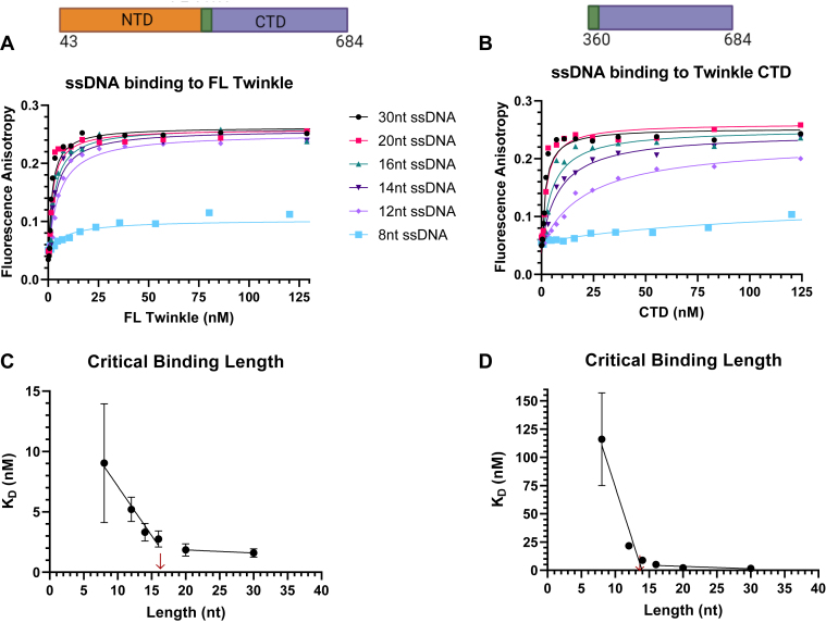
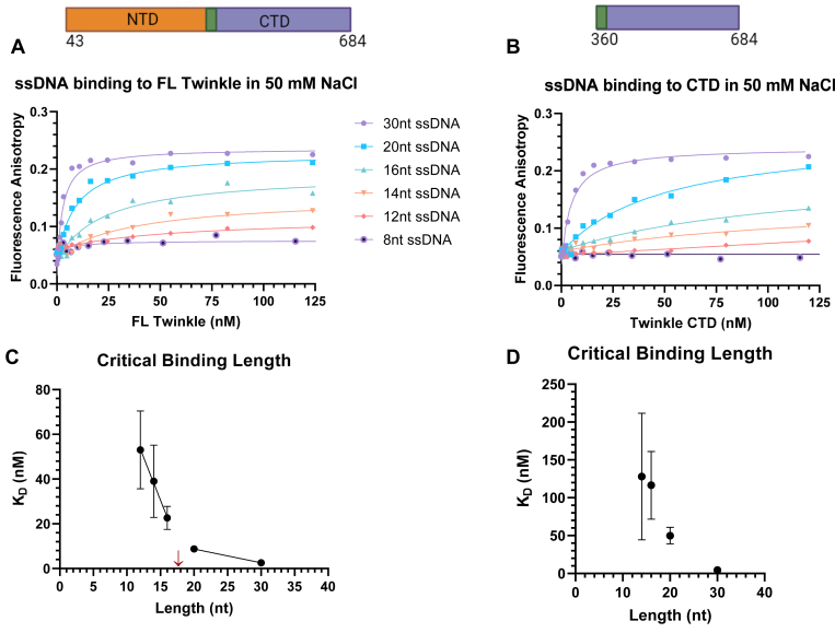
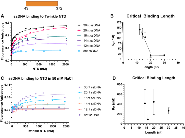
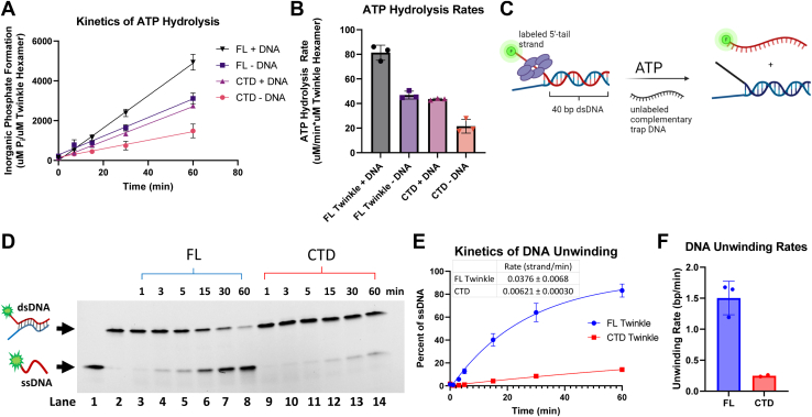
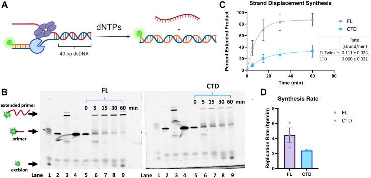
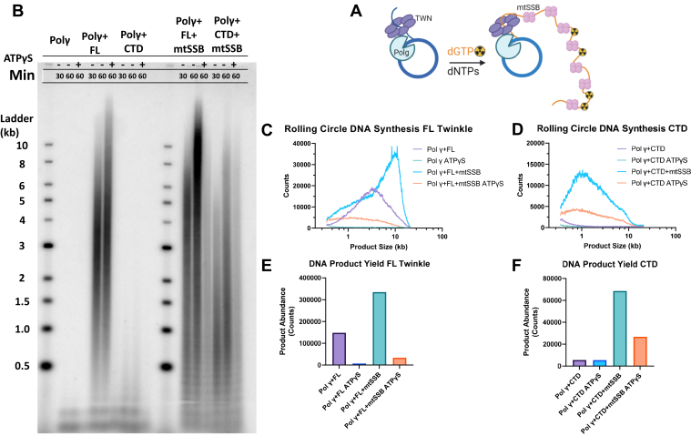
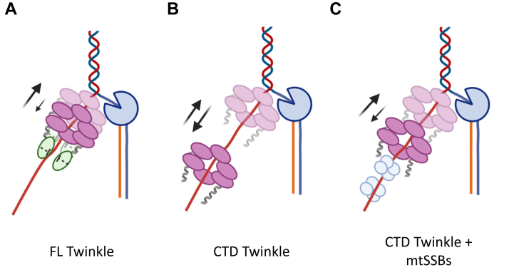
Twinkle is the ring-shaped replicative helicase within the human mitochondria with high homology to bacteriophage T7 gp4 helicase-primase. Unlike many orthologs of Twinkle, the N-terminal domain (NTD) of human Twinkle has lost its primase activity through evolutionarily acquired mutations. The NTD has no demonstrated activity thus far; its role has remained unclear. Here, we biochemically characterize the isolated NTD and C-terminal domain (CTD) with linker to decipher their contributions to full-length Twinkle activities. This novel CTD construct hydrolyzes ATP, has weak DNA unwinding activity, and assists DNA polymerase γ (Polγ)-catalyzed strand-displacement synthesis on short replication forks. However, CTD fails to promote multikilobase length product formation by Polγ in rolling-circle DNA synthesis. Thus, CTD retains all the motor functions but struggles to implement them for processive translocation. We show that NTD has DNA-binding activity, and its presence stabilizes Twinkle oligomerization. CTD oligomerizes on its own, but the loss of NTD results in heterogeneously sized oligomeric species. The CTD also exhibits weaker and salt-sensitive DNA binding compared with full-length Twinkle. Based on these results, we propose that NTD directly contributes to DNA binding and holds the DNA in place behind the central channel of the CTD like a "doorstop," preventing helicase slippages and sustaining processive unwinding. Consistent with this model, mitochondrial single-stranded DNA-binding protein (mtSSB) compensate for the NTD loss and partially restore kilobase length DNA synthesis by CTD and Polγ. The implications of our studies are foundational for understanding the mechanisms of disease-causing Twinkle mutants that lie in the NTD.

Keywords: DNA polymerase; Twinkle; helicase; mitochondria; mitochondrial diseases; replication; replisome.


Conflict of interest statement

Conflict of interest The authors declare that they have no conflicts of interest with the contents of this article.

Figures

Figure 1
Figure 1
Domain structure of the Twinkle subunit and its organization into a ring.A, the AlphaFold predicted structure for human Twinkle with the NTD in orange, the CTD in purple, and the linker region in green. Deletion position residues 360 and 372 are rendered as spheres and marked with arrows. B, the isolated subunit of the heptameric Twinkle (Protein Data Bank ID: 7T8C). C, cryo-EM structure of the heptameric Twinkle ring (Protein Data Bank ID: 7T8C). The primary structure schematic of the full-length construct with included amino acid residues is displayed above with the domains color coded. CTD, C-terminal domain; NTD, N-terminal domain.
Figure 2
Figure 2
Gel-filtration analysis to assess the oligomerization of purified FL Twinkle, CTD, and NTD constructs. Samples were injected in Superdex 200 Increase 10/300 GL Cytiva column with respective running at 0.3 ml/min and peak elution volume monitored via 280 nm absorbance. A, calibration curve shows the elution volumes of the Bio-Rad gel filtration protein standards as black squares, and the elution volume of each Twinkle construct is displayed as appropriately colored circles. B, FL human Twinkle elution profile monitored using absorbance at 280 nm. Expected locations of hexameric and monomeric FL Twinkle are marked with arrows as determined by the calibration curve, major peak elutes ∼heptamer. C, Twinkle CTD elution profile with the expected locations of hexameric and monomeric CTD marked with arrows, peak elution correlates to a dodecamer. D, Twinkle NTD elution profile with the expected locations of hexameric and monomeric NTD marked with arrows, peak elution corresponds to a monomer. The oligomerization state(s) most likely present in each spectrum based on the elution profile is displayed to the right, whereas the primary structure schematic of each construct with included amino acid residues is displayed above each respective graph. CTD, C-terminal domain; FL, full length; NTD, N-terminal domain.
Figure 3
Figure 3
Length-dependent binding of ssDNAs to FL Twinkle and CTD. The fluorescence anisotropy–based titrations were carried out in buffer containing 50 mM Tris acetate (pH 7.5), 10% glycerol, 0.05% Tween-20, and 0.5 mM DTT using 2.5 nM ssDNA (8, 12, 14, 16, 20, and 30 nt) titrated with hexameric concentrations of FL Twinkle and CTD. A, length dependence binding titration curves for FL Twinkle on a series of ssDNA lengths. B, length dependence binding titration curves for CTD on a series of ssDNAs. The equilibrium dissociation constant (KD) versus ssDNA length for FL Twinkle (C) and CTD (D). The primary structure schematic of each construct with included amino acid residues is displayed above each respective binding titration graph. Each curve fit to one-site binding hyperbolic equation and standard error of fit are shown. CTD, C-terminal domain; FL, full length.
Figure 4
Figure 4
ssDNA binding to FL Twinkle and CTD in 50 mM NaCl. The fluorescence anisotropy–based titrations were carried out in same buffer as for Figure 3 with 50 mM NaCl added. Each curve was fit to one-site binding hyperbolic equation. A, length dependence binding titration curves for FL Twinkle for the series of ssDNA lengths. B, length dependence binding titration curves for CTD for the series of ssDNAs. The equilibrium dissociation constant (KD) versus ssDNA length for FL Twinkle (C) and CTD (D). The primary structure schematic of each construct with included amino acid residues is displayed above each respective binding titration graph. Each curve fit to one-site binding hyperbolic equation and standard error of fit are shown. CTD, C-terminal domain; FL, full length.
Figure 5
Figure 5
Length-dependent binding of ssDNAs to Twinkle NTD. The fluorescence anisotropy–based titrations were carried out as detailed for Figures 3 and 4. A, DNA-binding titration curves of NTD for a series of ssDNA in no salt added conditions. B, NTD KDversus ssDNA length in no salt added conditions. C, DNA-binding curves for NTD in 50 mM NaCl conditions. D, NTD KDversus ssDNA length in 50 mM NaCl conditions. Each curve fit to one-site binding hyperbolic equation except for 30 and 20 nt ssDNA in no salt added conditions, which were fit to two-site binding hyperbolic equation, and standard error of fit are shown. NTD, N-terminal domain.
Figure 6
Figure 6
ATP hydrolysis and DNA unwinding kinetics of CTD and FL Twinkle.A, time courses of ATP hydrolysis reaction for CTD and FL Twinkle with and without DNA. ATP hydrolysis was measured using 30 nM CTD or FL Twinkle (hexamer) with and without 2.5 nM M13 ssDNA molecules in 8 mM magnesium acetate and 1 mM ATP spiked with [γ-32P] ATP. B, the ATP hydrolysis rates from three replicates from A are shown with the standard deviations. C, schematics of the unwinding reaction showing 5′-ssDNA tail–bound Twinkle catalyzing the release of fluorescent 5′-tail DNA from a 40-bp duplex in the presence of ATP and trap DNA. The unwinding assay was carried out using the same buffer with 10 nM forked DNA, 4.5 mM ATP, 8 mM magnesium acetate, 55.5 nM Twinkle hexamer, and 280 nM trap DNA. D, representative image of a 4 to 20% native polyacrylamide gel showing the time course of ssDNA formation by FL Twinkle and CTD. Lane 1: free 5′-tail ssDNA, lane 2: free forked DNA, and lanes 3 to 8 and lanes 9 to 14 show time courses of forked DNA unwinding by FL Twinkle and CTD, respectively. E, DNA unwinding kinetics showing proportion of 5′-tail release for the FL Twinkle and CTD fit to a one-phase exponential equation, rate displayed. F, the rate of DNA unwinding in base pairs/minute was calculated by multiplying the rate of strands unwound per minute by the number of base pairs in the fork, graphed with individual replicates as points and error bars representing SD. CTD, C-terminal domain; FL, full length.
Figure 7
Figure 7
Strand displacement DNA synthesis activity of Polγ with and without FL Twinkle or CTD.A, schematic shows the strand displacement DNA synthesis assay with fluorescein-labeled primer to monitor primer extension by the denaturing gel assay. Reactions were carried out with 300 nM Twinkle hexamer or CTD hexamer, 100 nM forked DNA, 200 nM Polγ, 300 μM dCTP, 100 μM remaining dNTPs, and 4 mM ATP. B, representative image of the 15% TBE–urea gel resolving the starting primer and extended primer from DNA synthesis. Samples from reactions with FL Twinkle or CTD were loaded in identical lanes in the two gels. Lane 1: fork DNA + Polγ reaction, lane 2: forked DNA alone, lane 3: Polγ + primer-template reaction, lane 4: free primer, and lanes 5 to 9: reactions with Polγ and FL Twinkle or CTD. C, proportion of the extended primer with Polγ and FL Twinkle or CTD (error bars represent three replicates). The kinetics fit to a single exponential equation to provide a composite rate of DNA synthesis over the 40-bp duplex (solid lines). D, rates of DNA synthesis in base pairs/minute, calculated by multiplying the rate of DNA strand synthesis per minute by the number of base pairs in the fork, for FL Twinkle and CTD with individual replicates as points and error bars representing standard deviation. CTD, C-terminal domain; FL, full length; Polγ, DNA polymerase γ; TBE, Tris–borate–EDTA.
Figure 8
Figure 8
Rolling circle DNA synthesis on the 70-bp minicircle fork DNA.A, schematic shows rolling circle DNA synthesis on the 70-bp minicircle forked DNA with Twinkle, Polγ, and mtSSB. Reactions were carried out using 20 nM FL Twinkle or CTD hexamer, 20 nM wildtype Polγ, 250 nM mtSSB when added, 10 nM 70 bp minicircle forked DNA, 2 mM ATP, 250 μM dNTPs, 25 μM dGTP spiked with [α-32P]dGTP at 37 °C. B, representative image of an 0.8% alkaline agarose gel showing the DNA products from the rolling circle DNA synthesis with the DNA size marker ladder. C, quantified counts of the 60 min reaction as a function of product length with FL Twinkle and Polγ with and without mtSSB and ATPγS. Product lengths were determined from the calibration curve generated from the DNA ladder run in the same gel. D, quantified counts of the 60 min reaction as a function of product length with CTD and Polγ with and without mtSSB and ATPγS. E, total DNA product counts for each reaction containing FL Twinkle and Polγ with and without mtSSB and ATPγS. F, the total product counts for each reaction containing CTD and Polγ with and without mtSSB and ATPγS. CTD, C-terminal domain; FL, full length; mtSSB, mitochondrial single-stranded DNA-binding protein; Polγ, DNA polymerase γ.
Figure 9
Figure 9
Proposed doorstop mechanism for the role of NTD in supporting processive translocation for DNA unwinding as part of a replisome with Polγ and mtSSBs.A, the NTD binding to the DNA emerging from the central channel of the CTD domains in FL Twinkle minimize backward motion (down arrow) promoting forward motion (up arrow). B, the CTD at the replication fork has more significant backward motion. C, the mtSSB molecules binding the DNA behind the CTD ring prevent excessive backward movements. CTD, C-terminal domain; FL, full length; mtSSB, mitochondrial single-stranded DNA-binding protein; NTD, N-terminal domain; Polγ, DNA polymerase γ.

Similar articles

Cited by

References

    1. Spelbrink J.N., Li F.Y., Tiranti V., Nikali K., Yuan Q.P., Tariq M., et al. Human mitochondrial DNA deletions associated with mutations in the gene encoding Twinkle, a phage T7 gene 4-like protein localized in mitochondria. Nat. Genet. 2001;28:223–231. - PubMed
    1. Fratter C., Gorman G.S., Stewart J.D., Buddles M., Smith C., Evans J., et al. The clinical, histochemical, and molecular spectrum of PEO1 (Twinkle)-linked adPEO. Neurology. 2010;74:1619–1626. - PMC - PubMed
    1. Remtulla S., Emilie Nguyen C.T., Prasad C., Campbell C. Twinkle-associated mitochondrial DNA depletion. Pediatr. Neurol. 2019;90:61–65. - PubMed
    1. Pierce S.B., Gulsuner S., Stapleton G.A., Walsh T., Lee M.K., Mandell J.B., et al. Infantile onset spinocerebellar ataxia caused by compound heterozygosity for Twinkle mutations and modeling of Twinkle mutations causing recessive disease. Cold Spring Harb Mol. Case Stud. 2016;2:a001107. - PMC - PubMed
    1. Percetti M., Franco G., Monfrini E., Caporali L., Minardi R., La Morgia C., et al. TWNK in Parkinson's disease: a movement disorder and mitochondrial disease center perspective study. Mov. Disord. 2022;37:1938–1943. - PMC - PubMed

Publication types