. 2022 Dec 17;19(1):305.
doi: 10.1186/s12974-022-02664-y.

Saturated very long-chain fatty acids regulate macrophage plasticity and invasiveness

Affiliations

Saturated very long-chain fatty acids regulate macrophage plasticity and invasiveness

Bettina Zierfuss et al. J Neuroinflammation. .

Abstract

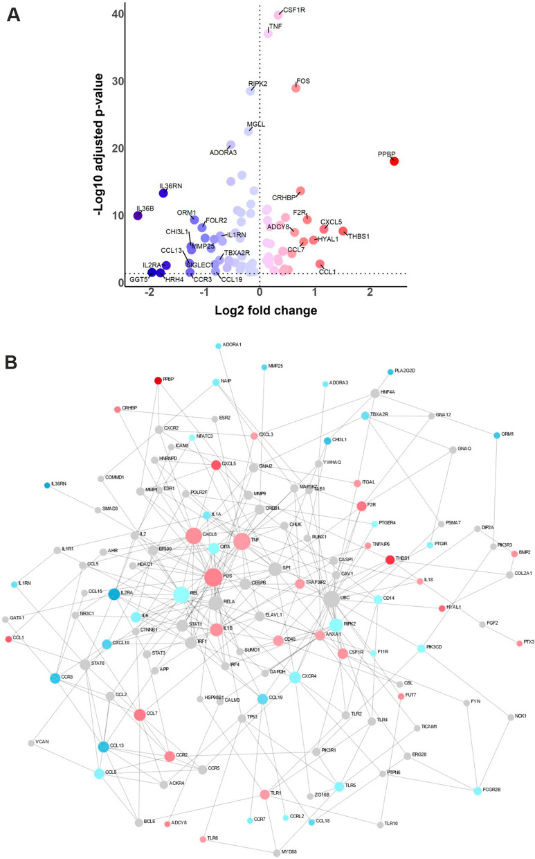
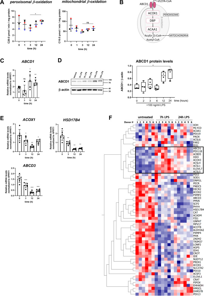
Saturated very long-chain fatty acids (VLCFA, ≥ C22), enriched in brain myelin and innate immune cells, accumulate in X-linked adrenoleukodystrophy (X-ALD) due to inherited dysfunction of the peroxisomal VLCFA transporter ABCD1. In its severest form, X-ALD causes cerebral myelin destruction with infiltration of pro-inflammatory skewed monocytes/macrophages. How VLCFA levels relate to macrophage activation is unclear. Here, whole transcriptome sequencing of X-ALD macrophages indicated that VLCFAs prime human macrophage membranes for inflammation and increased expression of factors involved in chemotaxis and invasion. When added externally to mimic lipid release in demyelinating X-ALD lesions, VLCFAs did not activate toll-like receptors in primary macrophages. In contrast, VLCFAs provoked pro-inflammatory responses through scavenger receptor CD36-mediated uptake, cumulating in JNK signalling and expression of matrix-degrading enzymes and chemokine release. Following pro-inflammatory LPS activation, VLCFA levels increased also in healthy macrophages. With the onset of the resolution, VLCFAs were rapidly cleared in control macrophages by increased peroxisomal VLCFA degradation through liver-X-receptor mediated upregulation of ABCD1. ABCD1 deficiency impaired VLCFA homeostasis and prolonged pro-inflammatory gene expression upon LPS treatment. Our study uncovers a pivotal role for ABCD1, a protein linked to neuroinflammation, and associated peroxisomal VLCFA degradation in regulating macrophage plasticity.

Keywords: Extracellular matrix degradation; Immune response; Lipid metabolism; Neuroinflammation; X-linked adrenoleukodystrophy.


Conflict of interest statement

The authors have no competing interests.

Figures

Fig. 1
Fig. 1
RNA-seq transcriptional profiling of X-ALD macrophages reveals alterations in genes associated with the immune response. A Volcano plot depicting differentially expressed genes belonging to the inflammatory response Gene Ontology Term (GO:0006954) of macrophages, derived by in vitro differentiation of monocytes from X-ALD patients compared to healthy controls (n = 9 for each). Red-coloured dots represent upregulated genes, whereas downregulated genes are indicated in blue. The fold change and the adjusted p-values are indicated on a log2 and log10 scale, respectively. Differentially expressed genes with either log2 fold changes higher than ± 0.6 or − log10 adjusted p-values higher than 18 were tagged with the indicated gene symbol. B Interactome graph of the inflammatory response Gene Ontology Term (GO:0006954) of macrophages derived from X-ALD patients compared to healthy controls (n = 9 for each). Nodes depict genes, while edges show protein–protein interactions between gene products. The colour of nodes depicts log2 fold change, with red indicating upregulated, blue downregulated and grey non-differentially expressed genes. The frequency of protein–protein interaction is reflected by the size of the node
Fig. 2
Fig. 2
The saturated VLCFA C26:0 activates the JNK stress kinase pathway but not TLR-mediated NFκB signalling. A Reporter Jurkat E6-1-NFκB::eGFP-TLR2/1, Jurkat E6-1-NFκB::eGFP-TLR2/6 and Jurkat E6-1-NFκB::eGFP-TLR4/CD14 cells, as well as reporter THP-1 E6-1-NFκB::eGFP-TLR4/CD14 cells were incubated with either the cognate TLR ligands flagellin, LPS, PAM3CSK4 or MALP2 or saturated LCFA (C16:0), saturated VLCFAs (C24:0, C26:0) or mono-unsaturated LCFA (C18:1) as indicated. After 24 h, eGFP expression was assessed by flow cytometry. The heatmap represents mean fold change to vehicle of 2 replicates. BG Primary human macrophages derived from healthy control donors (n = 2–4) were treated with either the solvent ethanol (vehicle), C16:0 (100 µM), C26:0 (100 µM) or LPS (100 ng/ml) for the indicated time. Immunoblotting was performed on cell lysates analysing the levels of BD phosphorylated and total NFκB-p65 or EG phosphorylated and total JNK1 (46 kDa)/JNK2 (55 kDa). H Macrophages derived from 3 healthy control donors were incubated with either C26:0 (100 µM), the CD36 inhibitor (CD36i) sulfosuccinimidyl oleate (100 µM) or both compounds for 24 h prior to immunoblotting for phosphorylated and total JNK1/JNK2. Values are shown as the mean fold change to vehicle control and error bars indicate standard deviation. One-way ANOVA and Fisher’s LSD multiple comparison test were performed in BG. Paired two-way Student’s t-test was performed in H. *p < 0.05; **p < 0.01; ***p < 0.001; ns not significant
Fig. 3
Fig. 3
Saturated VLCFA exposure initiates pro-inflammatory chemokine production in human macrophages. A Human macrophages derived from healthy control donors (n = 14) were treated with different concentrations of C26:0 or LPS for 24 h before expression of the pro-inflammatory cell surface marker CD86 was assessed by flow cytometry. B Human healthy control macrophages (n = 4–6) were treated with C26:0 (100 µM) or the solvent EtOH for the indicated time. RT-qPCR was carried out to measure mRNA levels of CXCL8, CCL3 and CCL4, and normalized to HPRT1. C Healthy control macrophages (n = 4) were treated with C26:0 (100 µM) or the solvent EtOH for 24 h. Supernatants were analysed for CXCL8, CCL3 and CCL4 protein levels using Luminex ELISA bead assays. The heat map indicates fold changes to solvent-treated samples. D RT-qPCR of CXCL8, CCL3 and CCL4 mRNA levels were normalized to HPRT1 in macrophages (n = 4) treated with either C16:0 (100 µM) or C26:0 (100 µM) for 12 or 24 h. E RT-qPCR of CXCL8 expression in healthy control macrophages (n = 3) treated with C26:0 (100 µM) or the CD36 inhibitor (CD36i) sulfosuccinimidyl oleate (100 µM) or with both compounds for 24 h. F RT-qPCR of MMP9, MMP14 and PLAUR normalized to HPRT1 was performed in healthy control macrophages (n = 5) treated with either C16:0 (100 µM) or C26:0 (100 µM) for 24 h. G Healthy control macrophages derived from 4 donors were treated with C26:0 (100 µM) or the solvent EtOH for 24 h before podosome structures were visualized by staining f-actin with AlexaFluor488-phalloidin and the frequency of podosome-positive macrophages being determined by fluorescence microscopy. One-way ANOVA and Fisher’s LSD multiple comparison test were performed in A and the ratio paired t-test was used in B, DG: **p < 0.01; *p < 0.05; ns not significant. Boxplots indicate median ± interquartile range, while whiskers show minimum and maximum. Bar graphs show means ± standard deviations
Fig. 4
Fig. 4
Macrophages modulate their saturated VLCFA content according to their activation state. A The levels of saturated and mono-unsaturated LCFAs and VLCFAs, as well as PUFAs of human macrophages incubated with LPS for 1, 3, 12 or 24 h were determined by ESI–MS. The heatmap represents mean values of macrophages derived from 5 healthy donors. RT-qPCR analysis of healthy control macrophages incubated with LPS for 1, 3, 12 or 24 h shows gene expression of B acute pro-inflammatory markers (TNFA, IL12B) as well as C enzymes involved in fatty acid synthesis (FASN, FADS2, SCD1, ELOVL1, ELOVL7). Data were normalized to HPRT1 and RACK1 or HPRT1 mRNA levels. D Macrophages derived from X-ALD patients (n = 5–7) and healthy controls (n = 5–7) were incubated with LPS for 24 h. RT-qPCR was carried out to assess expression of pro-inflammatory markers (IL1B, IL12B, IL6, CCL2 and CXCL8) and enzymes involved in fatty acid synthesis (FADS2, SCD1 and ELOVL7). For statistical analysis one-way ANOVA and Fisher’s LSD multiple comparison test were performed in B, C and the Mann–Whitney test was used in D: ***p < 0.001; **p < 0.01; *p < 0.05; ns not significant. Boxplots indicate median ± interquartile range, while whiskers show minimum and maximum. Bar graphs show means ± standard deviations
Fig. 5
Fig. 5
Saturated VLCFAs are degraded by peroxisomal β-oxidation with onset of pro-inflammatory resolution. Macrophages derived from healthy control donors were treated with LPS and incubated for the indicated time. A The mean values of C26:0 and C16:0 degradation by peroxisomal and mitochondrial β-oxidation normalized to protein content are shown (n = 3). B Scheme indicating the enzymes involved in peroxisomal β-oxidation (acyl-coenzyme A oxidase 1, ACOX1; D-bifunctional protein, DBP; acetyl-CoA acyltransferase 1, ACAA1). C RT-qPCR of ABCD1 normalized to HPRT1 mRNA levels (n = 6). D Immunoblot analysis to determine ABCD1 protein levels normalized to β-actin of macrophages derived from 4 healthy donors. Representative blot shows values from one healthy donor. E RT-qPCR of ACOX1, HSD17B4 and ABCD3 expression with normalization to HPRT1 mRNA levels (n = 6). F Microarray data from LPS-stimulated human primary macrophages (n = 6) were retrieved from Regan et al., (GSE85333) and log 2-fold changes are shown by the heatmap. One-way ANOVA and Fisher’s LSD comparison test was used for statistical analysis in A, CE. Bar graphs indicate means ± standard deviations. ***p < 0.001; **p < 0.01; *p < 0.05; ns not significant
Fig. 6
Fig. 6
The upregulation of ABCD1 in human macrophages entering pro-inflammatory resolution is mediated by LXRα. A, B Macrophages derived from healthy control donors were treated with LPS and incubated for the indicated time. The expression of NR1H3 (encoding LXRα) and CH25H normalized to HPRT1 was analysed by RT-qPCR (n = 6). C Macrophages from 4 healthy donors were treated with either 25-hydroxycholesterol (25-HC), the LXR antagonist GSK1440233 or the solvent EtOH for 24 h followed by analysis of ABCD1 expression by RT-qPCR. D Macrophages from 5 healthy donors were treated with the LXR-agonist T0901317 or solvent control for 24 h and ABCD1 levels normalized by HPRT1 were determined RT-qPCR. E Immunoblot analysis of macrophages treated with either 25-HC, the LXR-agonist T0901317 or solvent control to determine ABCD1 protein levels normalized to β-actin (n = 4). Representative blot of macrophages derived from one healthy donor is shown. F Macrophages derived from three healthy donors were treated with either LPS or LPS and the LXR antagonist GSK1440233 for 3 or 24 h before ABCD1 mRNA levels normalized for HPRT1 were determined by RT-qPCR. One-way ANOVA and Fisher’s LSD comparison test was used for statistical analysis in A and B. Ratio paired Student’s t test was performed on absolute values in CE and paired Student’s t test in F. ***p < 0.001; **p < 0.01; *p < 0.05; ns not significant
Fig. 7
Fig. 7
Proposed mechanism for how VLCFAs promote a pro-inflammatory and pro-invasive phenotype of human macrophages. In an acute pro-inflammatory response, macrophages react by increasing the levels of saturated VLCFAs to create a receptive environment in the plasma membrane that enables pro-inflammatory signalling and the production of factors required for invasion and adhesion. When applied externally to mimic the condition in acute cerebral X-ALD lesions, VLCFAs activate the CD36/JNK axis, thus also promoting a pro-inflammatory pro-invasive macrophage response culminating in the secretion of chemokines and matrix-degrading enzymes

References

    1. Hsieh W-Y, Zhou QD, York AG, Williams KJ, Scumpia PO, Kronenberger EB, et al. Toll-like receptors induce signal-specific reprogramming of the macrophage lipidome. Cell Metab. 2020;32(1):128–43. e5. doi: 10.1016/j.cmet.2020.05.003. - DOI - PMC - PubMed
    1. Dennis EA, Deems RA, Harkewicz R, Quehenberger O, Brown HA, Milne SB, et al. A mouse macrophage lipidome. J Biol Chem. 2010;285(51):39976–39985. doi: 10.1074/jbc.M110.182915. - DOI - PMC - PubMed
    1. Oishi Y, Spann NJ, Link VM, Muse ED, Strid T, Edillor C, et al. SREBP1 contributes to resolution of pro-inflammatory TLR4 signaling by reprogramming fatty acid metabolism. Cell Metab. 2017;25(2):412–427. doi: 10.1016/j.cmet.2016.11.009. - DOI - PMC - PubMed
    1. Kanoh H, Nitta T, Go S, Inamori K-I, Veillon L, Nihei W, et al. Homeostatic and pathogenic roles of GM3 ganglioside molecular species in TLR4 signaling in obesity. EMBO J. 2020;39(12):e101732. doi: 10.15252/embj.2019101732. - DOI - PMC - PubMed
    1. Turk BR, Theda C, Fatemi A, Moser AB. X-linked adrenoleukodystrophy: pathology, pathophysiology, diagnostic testing, newborn screening, and therapies. Int J Dev Neurosci. 2019;25:S0736-5748(19)30133-9. - PubMed

MeSH terms