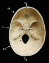
Fatal non-accidental pediatric cranial fracture risk and three-layered cranial architecture development
- PMID: 36529468
- PMCID: PMC10108079
- DOI: 10.1111/1556-4029.15183
Fatal non-accidental pediatric cranial fracture risk and three-layered cranial architecture development
Abstract
This study examines the influence of three-layered cranial architecture development upon blunt force trauma (BFT) cranial outcomes associated with pediatric non-accidental injury (NAI). Macroscopic and microscopic metric and morphological comparisons of subadult crania ranging from perinatal to 17 years of age chronicle the ontogenetic development and spatial and temporal variability in the emergence of a mature cranial architecture. Cranial vault thickness increases with subadult age, accelerating in the first 2 years of life due to rapid brain growth during this period. Three-layer differentiation of the cranial tables and diploë initiates by 3-6 months but is not consistently observed until 18 months to 2 years; diploë formation is not well developed until after age 4 and does not manifest a mature appearance until after age 8. These results allow topographic documentation of cortical and diploic development and temporal and spatial variability across the growing cranium. The lateral cranial vault is identified as expressing delayed development and reduced expression of the three-layer architecture, a pattern that continues into adulthood. Comparison of fracture locations from known BFT pediatric cases with identified cranial fracture high-risk impact regions shows a concordance and suggests the presence of a higher fracture risk associated with non-accidental BFT in the lateral vault region in subadults below the age of 2. The absence or lesser development of a three-layered architecture in subadults leaves their cranial bones, particularly in the lateral vault, thin and vulnerable to the effects of BFT.
Keywords: blunt force trauma; forensic anthropology; fracture risk; ontogenetic development; pediatric non-accidental injury; three-layer cranial architecture.
© 2022 The Authors. Journal of Forensic Sciences published by Wiley Periodicals LLC on behalf of American Academy of Forensic Sciences.
Figures


References
-
- Boyd D. The anatomical basis for fracture repair: recognition of the healing continuum and its forensic applications to investigations of pediatric and elderly abuse. In: Boyd CC, Boyd DC, editors. Forensic anthropology: theoretical framework and scientific basis. Chichester, UK: Wiley Publishing; 2018. p. 151–200.
-
- Gill JR, Andrew T, Gilliland MFG, Love J, Matshes E, Reichard RR. National Association of medical examiners position paper: recommendations for the postmortem assessment of suspected head trauma in infant and young children. Acad Forensic Pathol. 2013;4(2):206–13. 10.23907/2014.032 - DOI
-
- Duhaime A. Nonaccidental head injuries. In: Albright AL, Pollack IF, Adelson PD, editors. Principles and practice of pediatric neurosurgery. New York, NY: Thieme Medical Publishers Inc.; 2008. p. 791–802.
MeSH terms
LinkOut - more resources
Full Text Sources
Medical

