

Enhancement of Magnetic Surface-Enhanced Raman Scattering Detection by Tailoring Fe3O4@Au Nanorod Shell Thickness and Its Application in the On-site Detection of Antibiotics in Water
- PMID: 36530269
- PMCID: PMC9753213
- DOI: 10.1021/acsomega.2c06099
Enhancement of Magnetic Surface-Enhanced Raman Scattering Detection by Tailoring Fe3O4@Au Nanorod Shell Thickness and Its Application in the On-site Detection of Antibiotics in Water
Abstract
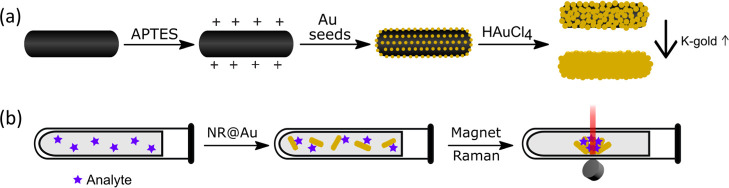
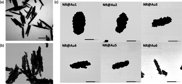
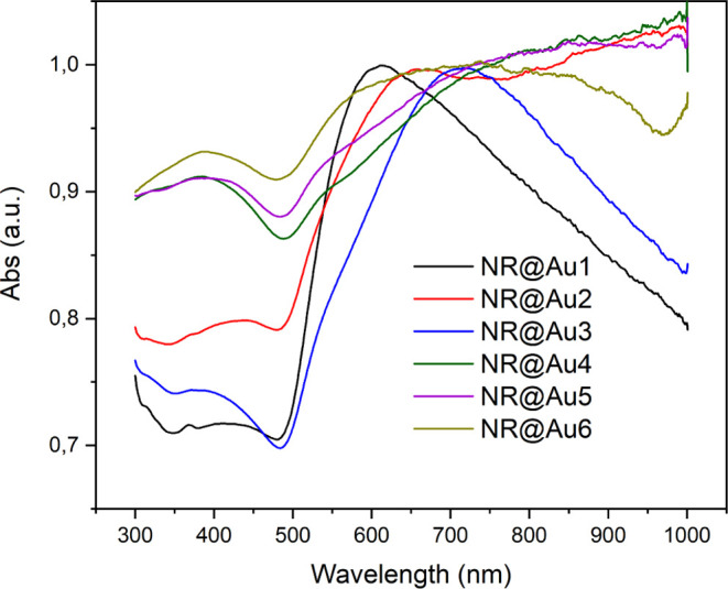
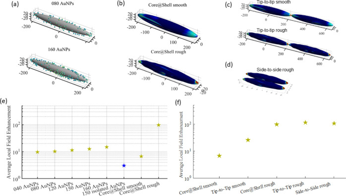
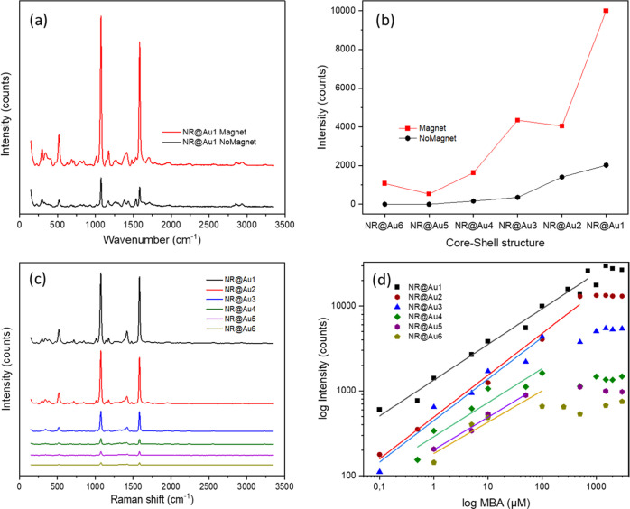
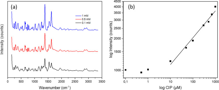
Surface-enhanced Raman scattering (SERS) has become a promising method for the detection of contaminants or biomolecules in aqueous media. The low interference of water, the unique spectral fingerprint, and the development of portable and handheld equipment for in situ measurements underpin its predominance among other spectroscopic techniques. Among the SERS nanoparticle substrates, those composed of plasmonic and magnetic components are prominent examples of versatility and efficiency. These substrates harness the ability to capture the target analyte, concentrate it, and generate unique hotspots for superior enhancement. Here, we have evaluated the use of gold-coated magnetite nanorods as a novel multifunctional magnetic-plasmonic SERS substrate. The nanostructures were synthesized starting from core-satellite structures. A series of variants with different degrees of Au coatings were then prepared by seed-mediated growth of gold, from core-satellite structures to core-shell with partial and complete shells. All of them were tested, using a portable Raman instrument, with the model molecule 4-mercaptobenzoic acid in colloidal suspension and after magnetic separation. Experimental results were compared with the boundary element method to establish the mechanism of Raman enhancement. The results show a quick magnetic separation of the nanoparticles and excellent Raman enhancement for all the nanoparticles both in dispersion and magnetically concentrated with limits of detection up to the nM range (∼50 nM) and a quantitative calibration curve. The nanostructures were then tested for the sensing of the antibiotic ciprofloxacin, highly relevant in preventing antibiotic contaminants in water reservoirs and drug monitoring, showing that ciprofloxacin can be detected using a portable Raman instrument at a concentration as low as 100 nM in a few minutes, which makes it highly relevant in practical point-of-care devices and in situ use.
© 2022 The Authors. Published by American Chemical Society.
Conflict of interest statement
The authors declare no competing financial interest.
Figures
References
-
- Managò S.; Tramontano C.; Delle Cave D.; Chianese G.; Zito G.; De Stefano L.; Terracciano M.; Lonardo E.; De Luca A. C.; Rea I. SERS Quantification of Galunisertib Delivery in Colorectal Cancer Cells by Plasmonic-Assisted Diatomite Nanoparticles. Small 2021, 17, 2101711.10.1002/smll.202101711. - DOI - PubMed
-
- Langer J.; Jimenez de Aberasturi D.; Aizpurua J.; Alvarez-Puebla R. A.; Auguié B.; Baumberg J. J.; Bazan G. C.; Bell S. E. J.; Boisen A.; Brolo A. G.; Choo J.; Cialla-May D.; Deckert V.; Fabris L.; Faulds K.; García de Abajo F. J.; Goodacre R.; Graham D.; Haes A. J.; Haynes C. L.; Huck C.; Itoh T.; Käll M.; Kneipp J.; Kotov N. A.; Kuang H.; Le Ru E. C.; Lee H. K.; Li J.-F.; Ling X. Y.; Maier S. A.; Mayerhöfer T.; Moskovits M.; Murakoshi K.; Nam J.-M.; Nie S.; Ozaki Y.; Pastoriza-Santos I.; Perez-Juste J.; Popp J.; Pucci A.; Reich S.; Ren B.; Schatz G. C.; Shegai T.; Schlücker S.; Tay L.-L.; Thomas K. G.; Tian Z.-Q.; Van Duyne R. P.; Vo-Dinh T.; Wang Y.; Willets K. A.; Xu C.; Xu H.; Xu Y.; Yamamoto Y. S.; Zhao B.; Liz-Marzán L. M. Present and Future of Surface-Enhanced Raman Scattering. ACS Nano 2020, 14, 28–117. 10.1021/acsnano.9b04224. - DOI - PMC - PubMed
LinkOut - more resources
Full Text Sources
Miscellaneous
