. 2022 Dec 2;7(49):44825-44835.
doi: 10.1021/acsomega.2c04911. eCollection 2022 Dec 13.

Antibiofilm Activity of PEGylated Branched Polyethylenimine

Affiliations

Antibiofilm Activity of PEGylated Branched Polyethylenimine

Hannah Panlilio et al. ACS Omega. .

Abstract

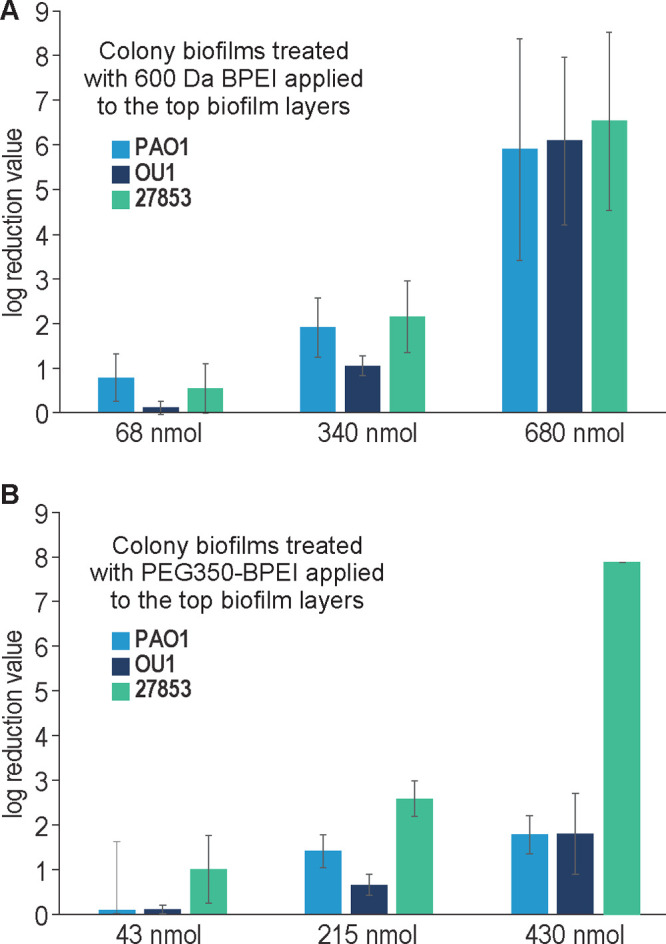
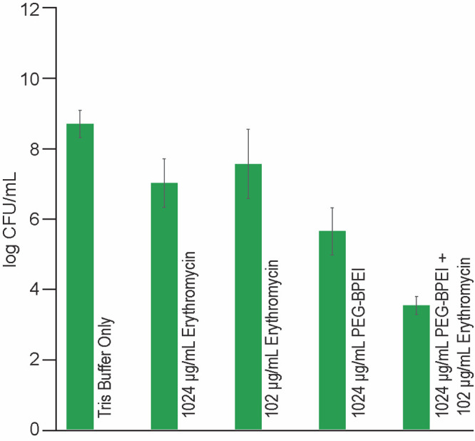
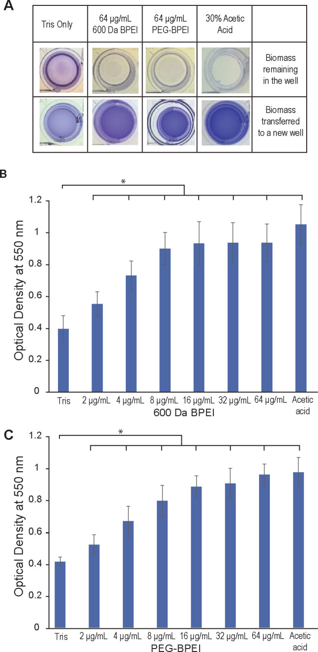
Biofilm formation is an adaptive resistance mechanism that pathogens employ to survive in the presence of antimicrobials. Pseudomonas aeruginosa is an infectious Gram-negative bacterium whose biofilm allows it to withstand antimicrobial attack and threaten human health. Chronic wound healing is often impeded by P. aeruginosa infections and the associated biofilms. Previous findings demonstrate that 600 Da branched polyethylenimine (BPEI) can restore β-lactam potency against P. aeruginosa and disrupt its biofilms. Toxicity concerns of 600 Da BPEI are mitigated by covalent linkage with low-molecular-weight polyethylene glycol (PEG), and, in this study, PEGylated BPEI (PEG350-BPEI) was found exhibit superior antibiofilm activity against P. aeruginosa. The antibiofilm activity of both 600 Da BPEI and its PEG derivative was characterized with fluorescence studies and microscopy imaging. We also describe a variation of the colony biofilm model that was employed to evaluate the biofilm disruption activity of BPEI and PEG-BPEI.


Conflict of interest statement

The authors declare no competing financial interest.

Figures

Figure 1
Figure 1
Illustration of the modified colony biofilm model. (A) Side profile of the biofilm colony grown on top of a porous membrane resting on TSA agar. (B) Treatment option (i) in a bulk solution of treatment agent. (C) Treatment option (ii) used cellulose filtration discs to deliver treatment to the biofilm surface and prevent the treatment solution from evaporating, followed by CFU counting.
Figure 2
Figure 2
Scanning electron micrographs of ATCC BAA-47 (PAO1) P. aeruginosa grown via colony biofilm model. Thick layers of P. aeruginosa cells are stacked on top of each other after 24 h (A). Closer inspection reveals a thick EPS surrounding the bacterial cells (B), encasing the bacterial cells. The SEM image in (A) shows a thick layer of rod-shaped P. aeruginosa cells embedded within the biomass. The biofilm cross-section shows layers of cells and EPS the comprise the biofilm (B). These SEM images confirm that P. aeruginosa cells are coated with EPS before treatments were added.
Figure 3
Figure 3
LRVs of 600 Da BPEI (A) and PEG350-BPEI (B). Error bars denote standard deviation (n = 5). Increasing nmol amounts of 600 Da BPEI delivered to the biofilms increases LRVs across different P. aeruginosa strains. LRVs of the highest concentration 600 Da BPEI are better than of PEG350-BPEI except for one strain: ATCC 27853.
Figure 4
Figure 4
Log CFU per mL of PAO1 suspension that survived after a single dose of each treatment in the ex vivo porcine skin model. Data set shows that before treatments, at time point zero (t = 0), there were ∼108 cells in the porcine skin. Buffer (Tris)-treated samples continued to grow more cells (∼109), and treatments with 600 Da BPEI and PEG350-BPEI caused a CFU reduction as observed in the colony biofilm model (n = 5; errors denote standard deviation. Data analysis indicates a significant difference between each of treatments, p-value <0.01).
Figure 5
Figure 5
Log CFU per mL of PAO1 suspension that survived after a single dose of each type of treatment in the ex vivo porcine skin model. Data set shows that even up to 1024 μg/mL of erythromycin only causes modest CFU/mL reduction (1.68 log CFU/mL) compared to buffer (Tris)-treated punches. Treatments with 102 μg/mL erythromycin alone or 1024 μg/mL PEG350-BPEI alone both caused 1.13 and 3.05 log CFU/mL, respectively. However, a combination of 1024 μg/mL PEG350-BPEI and 102 μg/mL erythromycin produced a 5.17 log CFU/mL reduction (n = 5; errors denote standard deviation. Data analysis indicates a significant difference between each of treatments, p-value <0.01)
Figure 6
Figure 6
Evaluation of biofilm biomass disruption via crystal violet staining. Biofilms of P. aeruginosa ATCC BAA-47 (PAO1) stained with crystal violet were treated with increasing concentrations of 600 Da BPEI (B) and PEG350-BPEI (C) for 3 h, in addition to negative and positive controls. The dissolved biofilms were transferred into a separate microtiter plate leaving remnants of undissolved biomass in the original microtiter plate (A). The mean OD550 nm of dissolved biofilms was measured and recorded. Error bars denote standard deviation (n = 12). OD550 nm readings show a significant difference between buffer-treated (Tris) control and treatments indicated with an asterisk (t test, p-value <0.01).
Figure 7
Figure 7
Biofilm disruption assay via crystal violet staining for P. aeruginosa BAA-47 (PAO1) and 27853 treated with 600 Da BPEI and PEG350-BPEI. The P. aeruginosa biofilms stained with crystal violet were treated with 600 Da BPEI and PEG350-BPEI for 3 h, in addition to negative and positive controls. The mean OD550 nm of dissolved biofilms was measured and recorded. Error bars denote standard deviation (n = 10). OD550 nm values indicate that PEGylated BPEI can disrupt P. aeruginosa with an activity similar to that of 600 Da BPEI (t test, p-value >0.01, denoted by not significant, n.s.). OD550 nm readings show significant difference between buffer-treated (Tris) control and treatments indicated with an asterisk (t test, p-value <0.01).
Figure 8
Figure 8
Fluorescence imaging of pyoverdine in P. aeruginosa. Buffer-treated control shows a layer of cells covered in a thin layer of EPS, resulting in a hazy film (A). A reduction of cells and EPS coating is observed when treated with 600-Da BPEI (B).
Figure 9
Figure 9
Scanning electron micrographs of ATCC BAA-47 (PAO1) P. aeruginosa biofilms after treatment via colony biofilm model. Buffer-treated (Tris) biofilms (A, C) are observed to have a film-like layer, whereas the 600 Da BPEI-treated biofilms lack this appearance (B, D). The shape and size of bacteria in 600 Da BPEI-treated biofilms are more resolved compared to the buffer-treated ones. Boxed regions in (A, )B are shown with higher magnification in (C, D).
Figure 10
Figure 10
ITC binding isotherm shows that 600 Da BPEI interacts with DNA. As 600 Da BPEI is injected into DNA solutions, initial binding events cause heat to be absorbed and cause DNA conformational reorganization followed by exothermic behavior as additional BPEI molecules binds to the phosphodiester backbone.

References

    1. Nussbaum S. R.; Carter M. J.; Fife C. E.; DaVanzo J.; Haught R.; Nusgart M.; Cartwright D. An Economic Evaluation of the Impact, Cost, and Medicare Policy Implications of Chronic Nonhealing Wounds. Value Health 2018, 21 (1), 27–32. 10.1016/j.jval.2017.07.007. - DOI - PubMed
    1. Bhargava A.; Hayakawa K.; Silverman E.; Haider S.; Alluri K. C.; Datla S.; Diviti S.; Kuchipudi V.; Muppavarapu K. S.; Lephart P. R.; Marchaim D.; Kaye K. S. Risk factors for colonization due to carbapenem-resistant Enterobacteriaceae among patients exposed to long-term acute care and acute care facilities. Infect Control Hosp Epidemiol 2014, 35 (4), 398–405. 10.1086/675614. - DOI - PubMed
    1. Marchaim D.; Chopra T.; Bogan C.; Bheemreddy S.; Sengstock D.; Jagarlamudi R.; Malani A.; Lemanek L.; Moshos J.; Lephart P. R.; Ku K.; Hasan A.; Lee J.; Khandker N.; Blunden C.; Geffert S. F.; Moody M.; Hiro R.; Wang Y.; Ahmad F.; Mohammadi T.; Faruque O.; Patel D.; Pogue J. M.; Hayakawa K.; Dhar S.; Kaye K. S. The burden of multidrug-resistant organisms on tertiary hospitals posed by patients with recent stays in long-term acute care facilities. Am. J. Infect Control 2012, 40 (8), 760–5. 10.1016/j.ajic.2011.09.011. - DOI - PubMed
    1. Henig O.; Cober E.; Richter S. S.; Perez F.; Salata R. A.; Kalayjian R. C.; Watkins R. R.; Marshall S.; Rudin S. D.; Domitrovic T. N.; Hujer A. M.; Hujer K. M.; Doi Y.; Evans S.; Fowler V. G. Jr.; Bonomo R. A.; van Duin D.; Kaye K. S. Antibacterial Resistance Leadership, G. A Prospective Observational Study of the Epidemiology, Management, and Outcomes of Skin and Soft Tissue Infections Due to Carbapenem-Resistant Enterobacteriaceae. Open Forum Infect Dis 2017, 4 (3), ofx157.10.1093/ofid/ofx157. - DOI - PMC - PubMed
    1. Ruffin M.; Brochiero E. Repair Process Impairment by Pseudomonas aeruginosa in Epithelial Tissues: Major Features and Potential Therapeutic Avenues. Front Cell Infect Microbiol 2019, 9 (182), 182.10.3389/fcimb.2019.00182. - DOI - PMC - PubMed

LinkOut - more resources