. 2022 Dec 1:12:1072579.
doi: 10.3389/fonc.2022.1072579. eCollection 2022.

A novel panel of clinically relevant miRNAs signature accurately differentiates oral cancer from normal mucosa

Affiliations

A novel panel of clinically relevant miRNAs signature accurately differentiates oral cancer from normal mucosa

Nikolay Mehterov et al. Front Oncol. .

Abstract

Introduction: Although a considerable body of knowledge has been accumulated regarding the early diagnosis and treatment of oral squamous cell carcinoma (OSCC), its survival rates have not improved over the last decades. Thus, deciphering the molecular mechanisms governing oral cancer will support the development of even better diagnostic and therapeutic strategies. Previous studies have linked aberrantly expressed microRNAs (miRNAs) with the development of OSCC.

Methods: We combined bioinformatical and molecular methods to identify miRNAs with possible clinical significance as biomarkers in OSCC. A set of 10 miRNAs were selected via an in silico approach by analysing the 3'untranslated regions (3'UTRs) of cancer-related mRNAs such as FLRT2, NTRK3, and SLC8A1, TFCP2L1 and etc. RT-qPCR was used to compare the expression of in silico identified miRNAs in OSCC and normal tissues (n=32).

Results: Among the screened miRNAs, miR-21-5p (p < 0.0001), miR-93-5p (p < 0.0197), miR-146b-5p (p <0.0012), miR-155-5p (p < 0.0001), miR-182-5p (p < 0.0001) were significantly overexpressed, whereas miR-133b (p < 0.05) was significantly downregulated in OSCC tissues, a scenario confirmed in two additional OSCC validation cohorts: Regina Elena National Cancer Institute (IRE cohort, N=74) and The Cancer Genome Atlas Data Portal (TCGA cohort, N=354). Initial stage tumors (T1, T2) expressed significantly higher levels of miR-133b (p < 0.0004) compared to more advanced ones (T3, T4). Also, we identified miR-93-5p (p < 0.0003), miR-133b (p < 0.0017) and miR-155-5p (p < 0.0004) as correlated with HPV-induced OSCC. The high expression of these 6 miRNAs as a signature predicted shorter disease-free survival (DFS) and could efficiently distinguish OSCC cases from healthy controls with areas under the curve (AUC) of 0.91 with sensitivity and specificity of 0.98 and 0.6, respectively. Further target identification analysis revealed enrichment of genes involved in FOXO, longevity, glycan biosynthesis and p53 cancer-related signaling pathways. Also, the selected targets were underexpressed in OSCC tissues and showed clinical significance related to overall survival (OS) and DFS.

Discussion: Our results demonstrate that a novel panel consisting of miR-21-5p, miR-93-5p, miR-133b, miR-146b-5p, miR-155-5p and miR-182-5p could be used as OSCC-specific molecular signature with diagnostic and prognostic significance related to OS and DFS.

Keywords: biomarker; disease-free survival; mRNA; miRNA; oral squamous cell carcinoma; overall survival.


Conflict of interest statement

The authors declare that the research was conducted in the absence of any commercial or financial relationships that could be construed as a potential conflict of interest.

Figures
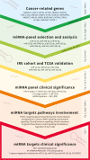
Figure 1
Figure 1
Experimental workflow used for the identification of clinically significant miRNAs and associated targets in OSCC.
Figure 2
Figure 2
miR-21-5p, miR-93-5p, miR-146b-5p, miR-155-5p, miR-182-5p and miR-133b are differentially expressed in three different OSCC patient cohorts. (A) Box plots of the normalized delta Ct expression of a 10 miRNA panel, evaluated in both OSCC and normal tissues from the UHSG cohort. Expression levels were determined by RT-qPCR and presented as fold differences. The reference control genes RNAU6 and SNORD72 were measured with two replicates in each PCR run, and the average Ct value was used for relative expression analysis. Relative miRNA abundance was calculated using the comparative 2-ΔΔCt method and normalized to geomean of the reference gene levels. Data are summarized from two technical replicates for each patient. N=normal, T=Tumoral. (B) Box plot showing the expression levels of miR-21-5p, miR-93-5p, miR-146b-5p, miR-155-5p, miR-182-5p, and miR-133b in normal, peritumoral and tumoral OSCC tissues from IRE cohort. N=normal, PT=Periumoral T=Tumoral. (C) Box plots showing expression levels of miR-21-5p, miR-93-5p, miR-146b-5p, miR-155-5p, miR-182-5p, and miR-133b in the tumoral and normal OSCC tissues from the TCGA cohort. P ≤ 0.05 was considered statistically significant for all assays. N=normal, T=HNSCC, Toc= OSCC.
Figure 3
Figure 3
miR-93-5p, miR-133b and miR-155-5p have HPV-dependent expression. Box plots showing the expression levels of miR-93-5p (A), miR-133b (B) and miR-155-5p (C) in both OSCC HPV+ and HPV- OSCC tissues from the TCGA cohort. P ≤ 0.05 was considered statistically significant for all assays.
Figure 4
Figure 4
miR-133b is predominantly expressed in initial stage tumors (T1, T2). Box plot showing the expression levels of miR-133b in initial (T1, T2) and advanced (T3, T4) stage tumors from the TCGA cohort. P ≤ 0.05 was considered as a statistically significant value.
Figure 5
Figure 5
High expression of the miRNA signature correlates with lower DFS and can distinguish OSCC from normal cases. (A) Kaplan–Meier analysis representing the correlation between expression levels of the miRNA signature and DFS in OSCC patients from the TCGA cohort. The TCGA cohort was divided in two subgroups according to high and low mean expression levels of each miRNA. High and low subgroups were established by evaluating positive and negative z-scores of the mean expression values of the miRNAs, respectively. For each KM curve, the hazard risk, confidence interval, and relative p-value (p) of the multivariate Cox analysis are also indicated. Cox regression was adjusted for T, N stage and HPV status. (B) Diagnostic ability of the miRNA signature for OSCC. ROC curve analysis of the miRNA signature in discriminating between patients and healthy individuals from the TCGA cohort.
Figure 6
Figure 6
miR-21-5p, miR-93-5p, miR-146b-5p, miR-155-5p, miR-182-5p and miR-133b control genes involved in FOXO, longevity, glycan biosynthesis and p53 pathways in OSCC. Bubble plot containing specific ontological groups of miR-21-5p, miR-93-5p, miR-146b-5p, miR-155-5p, miR-182-5p and miR-133b validated targets. The graphs represents only the GO groups above the established cut-off criteria (p with correction <0.05, minimal number of genes per group >10). Each bubble displays the number of differentially expressed genes assigned to the particular GO terms. The transparency of the bubbles shows the p-values (the darker the violet color, the closer to the border of p = 0.05).
Figure 7
Figure 7
Clinical utility of miR-21-5p, miR-93-5p, and miR-155-5p targets in OSCC. (A) Box-plot representing the expression of 14 genes negatively correlated with miR-21-5p, miR-93-5p, and miR-155-5p and differentially expressed between oral tumor tissues and their normal counterparts (P<0.05) in the TCGA OSCC cohort. Kaplan–Meier analysis representing the correlation between expression levels of miR-21-5p, miR-93-5p, and miR-155-5p targets considered as a group and OS (B) and DFS (C) in OSCC patients from the TCGA cohort. The TCGA cohort was divided in high and low groups, based on the positive and negative z-scores of the mean signal of the signature, respectively. The Hazard Ratio was assessed by multivariate cox regression analysis, adjusted for T,N stage and mutP53. For each KM curve, the hazard risk, confidence interval, and relative p-value (p) of the multivariate Cox analysis are also indicated.

References

    1. Nagao T, Warnakulasuriya S. Screening for oral cancer: Future prospects, research and policy development for Asia. Oral Oncol (2020) 105:104632. doi: 10.1016/j.oraloncology.2020.104632 - DOI - PubMed
    1. Gooi Z, Chan JY, Fakhry C. The epidemiology of the human papillomavirus related to oropharyngeal head and neck cancer. Laryngoscope (2016) 126(4):894–900. doi: 10.1002/lary.25767 - DOI - PubMed
    1. Berman TA, Schiller JT. Human papillomavirus in cervical cancer and oropharyngeal cancer: One cause, two diseases. Cancer (2017) 123(12):2219–29. doi: 10.1002/cncr.30588 - DOI - PubMed
    1. Gupta B, Johnson NW. Systematic review and meta-analysis of association of smokeless tobacco and of betel quid without tobacco with incidence of oral cancer in south Asia and the pacific. PLoS One (2014) 9(11):e113385. doi: 10.1371/journal.pone.0113385 - DOI - PMC - PubMed
    1. Montero PH, Patel SG. Cancer of the oral cavity. Surg Oncol Clin N Am (2015) 24(3):491–508. doi: 10.1016/j.soc.2015.03.006 - DOI - PMC - PubMed