

Assessment of the Classification of Age-Related Macular Degeneration Severity from the Northern Ireland Sensory Ageing Study Using a Measure of Dark Adaptation
- PMID: 36531574
- PMCID: PMC9754971
- DOI: 10.1016/j.xops.2022.100204
Assessment of the Classification of Age-Related Macular Degeneration Severity from the Northern Ireland Sensory Ageing Study Using a Measure of Dark Adaptation
Abstract
Purpose: To assess the differences in rod-mediated dark adaptation (RMDA) between different grades of age-related macular degeneration (AMD) severity using an OCT-based criterion compared with those of AMD severity using the Beckman color fundus photography (CFP)-based classification and to assess the association between the presence of subretinal drusenoid deposits (SDDs) and RMDA at different grades of AMD severity using an OCT-based classification.
Design: Cross-sectional study.
Participants: Participants from the Northern Ireland Sensory Ageing study (Queen's University Belfast).
Methods: Complete RMDA (rod-intercept time [RIT]) data, CFP, and spectral-domain OCT images were extracted. Participants were stratified into 4 Beckman groups (omitting late-stage AMD) and 3 OCT-based groups. The presence and stage of SDDs were identified using OCT.
Main outcome measures: Rod-intercept time data (age-corrected).
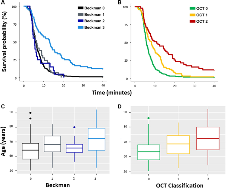
Results: Data from 459 participants (median [interquartile range] age, 65 [59-71] years) were stratified by both the classifications. Subretinal drusenoid deposits were detected in 109 eyes. The median (interquartile range) RMDA for the Beckman classification (Beckman 0-3, with 3 being intermediate age-related macular degeneration [iAMD]) groups was 6.0 (4.5-8.7), 6.6 (4.7-10.5), 5.7 (4.4-7.4), and 13.2 (6-21.1) minutes, respectively. OCT classifications OCT0-OCT2 yielded different median (interquartile range) values: 5.8 (4.5-8.5), 8.4 (5.2-13.3), and 11.1 (5.3-20.1) minutes, respectively. After correcting for age, eyes in Beckman 3 (iAMD) had statistically significantly worse RMDA than eyes in the other Beckman groups (P ≤ 0.005 for all), with no statistically significant differences between the other Beckman groups. Similarly, after age correction, eyes in OCT2 had worse RMDA than eyes in OCT0 (P ≤ 0.001) and OCT1 (P < 0.01); however, there was no statistically significant difference between eyes in OCT0 and eyes in OCT1 (P = 0.195). The presence of SDDs was associated with worse RMDA in OCT2 (P < 0.01) but not in OCT1 (P = 0.285).
Conclusions: Eyes with a structural definition of iAMD have delayed RMDA, regardless of whether a CFP- or OCT-based criterion is used. In this study, after correcting for age, the RMDA did not differ between groups of eyes defined to have early AMD or normal aging, regardless of the classification. The presence of SDDs has some effect on RMDA at different grades of AMD severity.
Keywords: AMD, age-related macular degeneration; AdaptDx; Age-related macular degeneration; Beckman; CFP, color fundus photography; OCT-based grading; RIT, rod-intercept time; RMDA, rod-mediated dark adaptation; RPE, retinal pigment epithelium; Rod-mediated dark adaptation; SD-OCT, spectral-domain OCT; SDD, subretinal drusenoid deposit; VA, visual acuity; iAMD, intermediate age-related macular degeneration.
© 2022 by the American Academy of Ophthalmology.
Figures


References
-
- Bird A.C., Bressler N.M., Bressler S.B., et al. An international classification and grading system for age-related maculopathy and age-related macular degeneration. Surv Ophthalmol. 1995;39:367–374. - PubMed
-
- Klein R., Davis M.D., Magli Y.L., et al. The Wisconsin age-related maculopathy grading system. Ophthalmology. 1991;98:1128–1134. - PubMed
Grants and funding
LinkOut - more resources
Full Text Sources
Miscellaneous
