Mitochondrial Ultrastructural Defects in NDUFS3-Related Disorder
- PMID: 36531773
- PMCID: PMC9757518
- DOI: 10.4103/jpn.JPN_182_20
Mitochondrial Ultrastructural Defects in NDUFS3-Related Disorder
Abstract
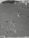
Complex I, the largest multisubunit enzyme complex of the respiratory chain, has a vital role in the energy production of the cell, and the clinical spectrum of complex I deficiency varies from severe lactic acidosis in infants to muscle weakness in adults. Pathogenic variants of NDUFS3 (constitutes the catalytic core of the complex I) have been reported in a small number of patients with variable phenotypes. We describe a girl with a history of infantile-onset nonepileptic myoclonus, who developed myopathy at the age of 2 years. Next-generation sequencing revealed compound heterozygous for two variants in the NDUSF3 gene. The electron-microscopic study of the skeletal muscle showed an increase in the number of mitochondria inside the myofibers; mitochondria were variably enlarged with some irregularity and were aligned perpendicular to the myofibrils in a stacked-up manner. This is the first description of mitochondrial ultrastructural abnormality in an individual with NDUFS3-related disorder.
Keywords: Complex I; NDUFS3; mitochondria; myoclonus; myopathy.
Copyright: © 2021 Journal of Pediatric Neurosciences.
Conflict of interest statement
The author declares no potential conflicts of interest with respect to the research, authorship, and publication of this article.
Figures


References
-
- Pagniez-Mammeri H, Lombes A, Brivet M, Ogier-de Baulny H, Landrieu P, Legrand A, et al. Rapid screening for nuclear genes mutations in isolated respiratory chain complex I defects. Mol Genet Metab. 2009;96:196–200. - PubMed
-
- Haack TB, Haberberger B, Frisch EM, Wieland T, Iuso A, Gorza M, et al. Molecular diagnosis in mitochondrial complex I deficiency using exome sequencing. J Med Genet. 2012;49:277–83. - PubMed
-
- Lou X, Shi H, Wen S, Li Y, Wei X, Xie J, et al. A novel NDUFS3 mutation in a Chinese patient with severe Leigh syndrome. J Hum Genet. 2018;63:1269–72. - PubMed
Publication types
LinkOut - more resources
Full Text Sources
Miscellaneous
