

Validating fatty acid binding protein 3 as a diagnostic and prognostic biomarker for peripheral arterial disease: A three-year prospective follow-up study
- PMID: 36531981
- PMCID: PMC9755058
- DOI: 10.1016/j.eclinm.2022.101766
Validating fatty acid binding protein 3 as a diagnostic and prognostic biomarker for peripheral arterial disease: A three-year prospective follow-up study
Abstract
Background: Patients with peripheral arterial disease (PAD) often remain undiagnosed and therefore suboptimally managed. Here, we investigated the diagnostic and prognostic potential of fatty acid binding protein 3 (FABP3) in patients with PAD.
Methods: In the discovery phase, 374 PAD and 184 non-PAD patients were recruited from vascular surgery ambulatory clinics at St. Michael's Hospital (Toronto, Ontario, Canada) between October 4, 2017 to October 29, 2018. The diagnostic ability of baseline FABP3 level was investigated through receiver operator characteristic (ROC) curves to determine two cutoff points: 1) an exclusionary "rule out" cutoff point, and 2) a confirmatory "rule in" cutoff point. Next, these cutoff points were confirmed in the external validation phase using a separate cohort of 312 patients (180 PAD and 132 non-PAD) recruited from ambulatory vascular surgery clinics at St. Michael's Hospital (Canada) between November 6, 2018-July 30, 2019. Cox regression analyses were used to explore the independent association between FABP3 and major adverse limb events (MALE - defined as need for arterial revascularization or major amputation) and decrease in ankle-brachial index (ABI -defined as drop ≥0.15) during 3 years of follow-up.
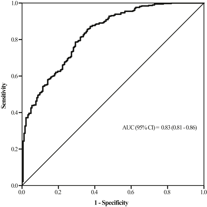
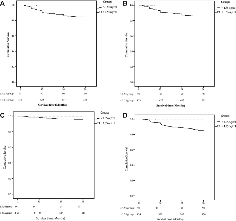
Findings: In the discovery phase, FABP3 levels were significantly elevated in patients with PAD compared to non-PAD patients. ROC analysis demonstrated that FABP3 had an AUC of 0.83 (95% CI: 0.81-0.86, p-value < 0.001). FABP3 exclusionary cutoff was <1.55 ng/ml (sensitivity = 96%; specificity = 40%), whereas FABP3 confirmatory cutoff was >3.55 ng/ml (sensitivity = 43%; specificity = 95%) - values that were confirmed in the external validation phase. Cox regression analysis demonstrated FABP3 to be an independent predictor of increase in MALE [HR = 1.14 (1.03-1.29); p-value = 0.010] and worsening PAD status (drop in ABI >0.15 [HR = 1.11 (1.02-1.19); p-value = 0.009]).
Interpretation: Our findings suggested that FABP3 levels can be used as both a diagnostic and prognostic biomarker for PAD, and may facilitate risk stratification in select individuals for purposes of vascular evaluation or intensive medical management.
Funding: Funding for this study was provided by the Bill and Vicky Blair Foundation.
Keywords: Biomarker; Diagnostic; FABP3; Fatty acid binding protein 3; Peripheral arterial disease; Prognostic.
© 2022 The Authors.
Conflict of interest statement
Dr. John Eikelboom reports consulting fees/honoraria and/or grant support from 10.13039/100004325AstraZeneca, Bayer Boehringer-Ingelheim, Bristol-Myer-Squibb/10.13039/100004319Pfizer, 10.13039/501100002973Daiichi-Sankyo, 10.13039/100004312Eli Lilly & Co, 10.13039/100004330GlaxoSmithKline, 10.13039/100004319Pfizer, 10.13039/100005565Janssen, 10.13039/100004339sanofi-aventis, 10.13039/501100011725Servier. The remaining authors have nothing to disclose.
Figures
References
-
- Fowkes F.G.R., Rudan D., Rudan I., et al. Comparison of global estimates of prevalence and risk factors for peripheral artery disease in 2000 and 2010: a systematic review and analysis. Lancet. 2013;382:1329–1340. - PubMed
-
- Weitz J.I., Byrne J., Clagett G.P., et al. Diagnosis and treatment of chronic arterial insufficiency of the lower extremities: a critical review. Circulation. 1996;94:3026–3049. - PubMed
-
- Hirsch A.T., Criqui M.H., Treat-Jacobson D., et al. Peripheral arterial disease detection, awareness, and treatment in primary care. JAMA. 2001;286:1317–1324. - PubMed
-
- Walsh D.B., Gilbertson J.J., Zwolak R.M., et al. The natural history of superficial femoral artery stenoses. J Vasc Surg. 1991;14:299–304. - PubMed
LinkOut - more resources
Full Text Sources
Miscellaneous
