Review
. 2022 Dec 1:13:1050478.
doi: 10.3389/fimmu.2022.1050478. eCollection 2022.

SARS-CoV-2 infection of phagocytic immune cells and COVID-19 pathology: Antibody-dependent as well as independent cell entry

Affiliations
Review

SARS-CoV-2 infection of phagocytic immune cells and COVID-19 pathology: Antibody-dependent as well as independent cell entry

Olga Matveeva et al. Front Immunol. .

Abstract

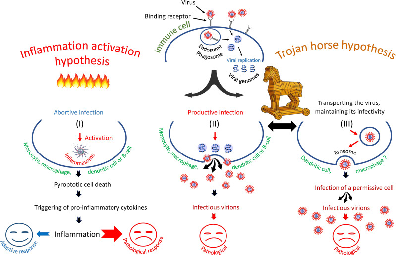
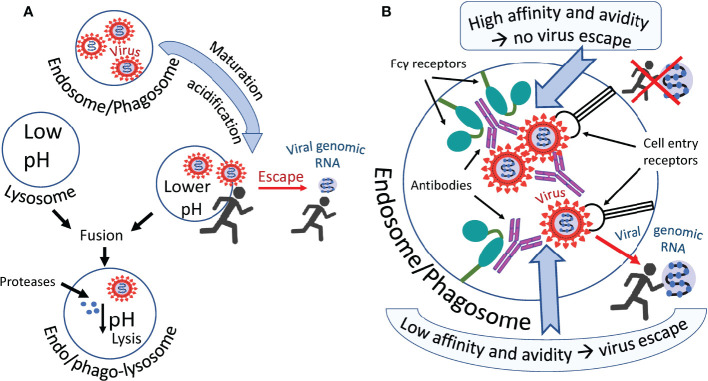
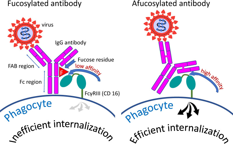
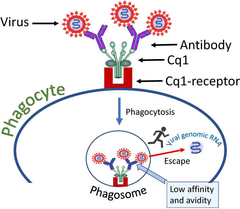
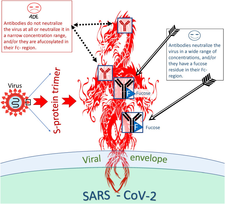
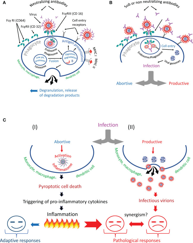
Our review summarizes the evidence that COVID-19 can be complicated by SARS-CoV-2 infection of immune cells. This evidence is widespread and accumulating at an increasing rate. Research teams from around the world, studying primary and established cell cultures, animal models, and analyzing autopsy material from COVID-19 deceased patients, are seeing the same thing, namely that some immune cells are infected or capable of being infected with the virus. Human cells most vulnerable to infection include both professional phagocytes, such as monocytes, macrophages, and dendritic cells, as well as nonprofessional phagocytes, such as B-cells. Convincing evidence has accumulated to suggest that the virus can infect monocytes and macrophages, while data on infection of dendritic cells and B-cells are still scarce. Viral infection of immune cells can occur directly through cell receptors, but it can also be mediated or enhanced by antibodies through the Fc gamma receptors of phagocytic cells. Antibody-dependent enhancement (ADE) most likely occurs during the primary encounter with the pathogen through the first COVID-19 infection rather than during the second encounter, which is characteristic of ADE caused by other viruses. Highly fucosylated antibodies of vaccinees seems to be incapable of causing ADE, whereas afucosylated antibodies of persons with acute primary infection or convalescents are capable. SARS-CoV-2 entry into immune cells can lead to an abortive infection followed by host cell pyroptosis, and a massive inflammatory cascade. This scenario has the most experimental evidence. Other scenarios are also possible, for which the evidence base is not yet as extensive, namely productive infection of immune cells or trans-infection of other non-immune permissive cells. The chance of a latent infection cannot be ruled out either.

Keywords: ADE; COVID-19 pathogenesis; SARS-CoV-2; immune cells infection; macrophage infection; monocyte infection; phagocyte infection; pyroptosis.


Conflict of interest statement

Authors OM and DL was employed by Sendai Viralytics, LLC. The remaining authors declare that the research was conducted in the absence of any commercial or financial relationships that could be construed as a potential conflict of interest.

Figures

Figure 1
Figure 1
Three scenarios of SARS-CoV-2 infection of immune cells: (I) entry of SARS-CoV-2 into immune cells triggers pyroptosis and inflammatory cascade, (II) entry of virus into cells leads to productive infection or, (III) entry of virus promotes its transfer to primary permissive target cells, causing their infection. The second and third scenarios are consistent with the "Trojan horse" hypothesis.
Figure 2
Figure 2
Hypothetical ways in which the virus escapes phagosomes. (A) The virus leaves the endosome/phagosome during its maturation and acidification. (B) Antibodies that firmly retain the virus in the immune complex most likely prevent it from leaving the organelle as shown in the upper part of the figure. Antibodies not firmly holding the virus allow it to escape from the organelle into the cytoplasm, as shown in the lower part of the figure. Perhaps acidification of the endosome/phagosome during its maturation reduces the affinity of some antibodies to the virus, promoting the exit of the virus from the immune complex and facilitating its exit from this organelle.
Figure 3
Figure 3
Distinction between fucosylated and afucosylated antibodies. Fucosylated antibodies have a lower affinity for the cellular receptor FcγRIIIa (CD16a) compared to afucosylated antibodies. Consequently, these antibodies contribute less to the internalization of the pathogen by cells possessing this receptor than do afucosylated antibodies.
Figure 4
Figure 4
Hypothetical mechanism of SARS-CoV-2 entry into a cell via complement receptors. The low affinity of the antibody, which causes a loose connection to the virus, allows the virus to escape the complex and fuse its envelope to the phagosome membrane, thus introducing its genome into the cell cytoplasm.
Figure 5
Figure 5
Distinction between antibodies that are more or less likely to cause ADE. Antibodies capable and incapable of causing ADEs differ both at the level of epitope recognition and by the type of antibody itself. Thus, antibodies to certain antigenic epitopes are more prone to cause ADEs than antibodies directed at other viral epitopes. In addition, afucosylated antibodies are more likely to cause ADEs compared to fucosylated antibodies. However, even antibodies capable of inducing ADE at one concentration can neutralize the virus at another and be protective against the virus.
Figure 6
Figure 6
Antibody-dependent phagocytosis of SARS-CoV-2 in norm and pathology. (A) The phagocyte destroys the internationalized pathogen through the phagolysosomal pathway and presents its antigens via the major histocompatibility complexes (MHC I and MHC II). (B) The phagocyte engulfs but cannot inactivate SARS-CoV-2 and becomes infected. It is likely that the infection occurs due to the synergistic action of cell entry receptors and Fc receptors. (C) The virus escapes from the endosome/phagosome and the escape results in (I) abortive or (II) productive viral infection.

Similar articles

Cited by

References

    1. Gordon S. Phagocytosis: An immunobiologic process. Immunity (2016) 44(3):463–75. doi: 10.1016/j.immuni.2016.02.026 - DOI - PubMed
    1. Yutin N, Wolf MY, Wolf YI, Koonin EV. The origins of phagocytosis and eukaryogenesis. Biol Direct (2009) 4:9. doi: 10.1186/1745-6150-4-9 - DOI - PMC - PubMed
    1. Mills DB. The origin of phagocytosis in earth history. Interface Focus (2020) 10(4):20200019. doi: 10.1098/rsfs.2020.0019 - DOI - PMC - PubMed
    1. Rosales C, Uribe-Querol E. Phagocytosis: A fundamental process in immunity. BioMed Res Int (2017) 2017:9042851. doi: 10.1155/2017/9042851 - DOI - PMC - PubMed
    1. Hortova-Kohoutkova M, Tidu F, De Zuani M, Sramek V, Helan M, Fric J. Phagocytosis-inflammation crosstalk in sepsis: New avenues for therapeutic intervention. Shock (2020) 54(5):606–14. doi: 10.1097/SHK.0000000000001541 - DOI - PMC - PubMed

Publication types

Substances