. 2022 Dec 2:13:1035556.
doi: 10.3389/fimmu.2022.1035556. eCollection 2022.

Kinetics of free and ligand-bound atacicept in human serum

Affiliations

Kinetics of free and ligand-bound atacicept in human serum

Mahya Eslami et al. Front Immunol. .

Abstract

BAFF (B cell activation factor of the TNF family/B lymphocyte stimulator, BLyS) and APRIL (a proliferation-inducing ligand) are targeted by atacicept, a decoy receptor consisting of the extracellular domain of TACI (transmembrane activator and calcium-modulator and cyclophilin (CAML) interactor) fused to the Fc portion of human IgG1. The purpose of the study was to characterize free and ligand-bound atacicept in humans. Total and active atacicept in serum of healthy volunteers receiving a single dose of subcutaneous atacicept or in patients treated weekly for one year were measured by ELISA, Western blot, or cell-based assays. Pharmacokinetics of free and bound atacicept were predicted based on total atacicept ELISA results. Persistence of complexes of purified atacicept bound to recombinant ligands was also monitored in mice. Results show that unbound or active atacicept in human serum exceeded 0.1 µg/ml for one week post administration, or throughout a 1-year treatment with weekly administrations. After a single administration of atacicept, endogenous BAFF bound to atacicept was detected after 8 h then increased about 100-fold within 2 to 4 weeks. Endogenous heteromers of BAFF and APRIL bound to atacicept also accumulated, but atacicept-APRIL complexes were not detected. In mice receiving intravenous injections of purified complexes pre-formed in vitro, atacicept-BAFF persisted longer (more than a week) than atacicept-APRIL (less than a day). Thus, only biologically inactive BAFF and BAFF-APRIL heteromers accumulate on atacicept in vivo. The measure of active atacicept provides further support for the once-weekly dosing regimen implemented in the clinical development of atacicept.

Trial registration: ClinicalTrials.gov NCT00624338.

Keywords: APRIL; BAFF; atacicept; heteromers; reporter cells.


Conflict of interest statement

PS was supported by a research grant from Merck Healthcare KGaA, Darmstadt, Germany. OP is employee of Merck Institute for Pharmacometrics, Lausanne, Switzerland, an affiliate of Merck KGaA. ÖY and DW were employee of Merck Healthcare KGaA, Darmstadt, Germany at the time of the study. OD was employed by Adipogen Life Sciences. The remaining authors declare that the research was conducted in the absence of any commercial or financial relationships that could be construed as a potential conflict of interest.

Figures

Figure 1
Figure 1
Measure of active atacicept in the serum samples from atacicept-injected subjects with a reporter cell-based assay. (A) Increasing concentrations of atacicept can block Fc-BAFF and prevent death of BAFFR : Fas reporter cells. (B) Linear relationship between the concentration of Fc-BAFF needed to kill half of BAFFR : Fas reporter cells and the concentration of atacicept. (C) Active atacicept concentration in subject’s serum is quantified be measuring the EC50 of Fc-BAFF on BAFFR : Fas reporter cells. (D–F) Same as panels (A–C), but using BCMA : Fas reporter cells and Fc-APRIL to measure the ability of active atacicept to inhibit APRIL.
Figure 2
Figure 2
Time versus concentration profiles for subjects receiving increasing doses of atacicept and corresponding Western blot analyses of serum samples. (A) Active and total atacicept levels were measured in serum samples of healthy subjects who received a single dose of atacicept (25, 75 or 150 mg). 0.5 µl of serum samples at different time points were also analyzed by denaturing (SDS) but non-reducing Western blot with an anti-TACI (Ata1) antibody. Superimposed graphs show concentrations of active (triangles) or total (circles) atacicept measured in the same samples. cplx: atacicept-containing complex. ata, atacicept. TD, treatment day. (B) Active atacicept as a function of time in sera of subjects receiving a single dose of 25 (opened circles), 75 (grey diamonds) or 150 mg (black triangles) atacicept (n=3 per dose. Mean ± SEM). The time interval corresponding to the first week post-administration is shown. (C) Same as panel B, but for total atacicept levels measured after acid dissociation of ligands in a validated atacicept ELISA. (D) Difference between logarithms of total versus active atacicept concentrations, as a function of time after administration. Non-parametric one-way ANOVA with Kruskal-Wallis’ multiple comparison test. ns, not significant, *p < 0.05. The same set of data was used for all panels. Western blot experiment performed once in this format, and once with TD 0, 4, 14, 28 and 42 for all subjects. Total atacicept measured once. Active atacicept measured twice for all subjects and time points, plus once for TD 0, 4, 14, 28 and 42.
Figure 3
Figure 3
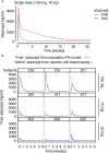
Prediction and measures of free or active atacicept in subjects receiving a single atacicept dose. (A) Predicted PK profiles for total and free atacicept for a typical 70 kg subject who received a single dose of 150 mg atacicept. Red line: total atacicept. Blue line: free atacicept. (B) “Active” atacicept PK profile (red dots) compared with model-derived “free” atacicept PK profiles (blue line) in nine subjects receiving the indicated doses of atacicept.
Figure 4
Figure 4
Prediction of free atacicept and measure of active atacicept in SLE patients receiving multiple injections of atacicept. (A) Population PK model for a 60 kg subject receiving 150 mg atacicept twice a week for 4 weeks, then weekly for 48 more weeks, with blue and red lines indicating predicted levels of free and total atacicept, respectively. (B) Active and total atacicept in 15 SLE patients (10 x 150 mg, 5 x 75 mg), before treatment, at weeks 4, 24 and 52 of treatment, and 12 weeks after treatment end (FU 12 W). Two ways ANOVA with Sidák’s multiple comparison test was used to compare active versus total atacicept at each time point. ns, not significant. *p < 0.05. Experiment performed once. (C) Active atacicept measured pre-dose, at the indicated time points during a 1-year atacicept treatment with bi-weekly administration of 150 mg atacicept for 4 weeks, then weekly for 48 further weeks, and at follow-up 12 weeks after treatment end (FU 12 W), in seven SLE patients. Serum samples (1 µl) collected at trough were analyzed in parallel by denaturing, but non-reducing SDS-PAGE followed by Western blot with anti-TACI mAb Ata1. cplx: atacicept-containing complex. ata: atacicept. NHS, normal human serum. Experiment was performed once in this format, and twice with reduced numbers of patients and time points.
Figure 5
Figure 5
Serum levels of endogenous BAFF and BAFF-APRIL heteromers increase in a time-dependent manner after administration of atacicept. (A) hBAFF ELISA signals upon titration of a BAFF standard alone or in the presence of the indicated fixed concentrations of atacicept. This experiment was performed twice under slightly different conditions (including Figure 6 ). (B) Healthy subjects received a single dose of atacicept at either 25 (black symbols), 75 (grey symbols) or 150 mg (opened symbols). BAFF levels were quantified in serum samples by ELISA in the presence of an additional saturating level of 3 μg/ml of atacicept. This experiment was performed twice. (C) Endogenous BAFF and APRIL levels were measured by ELISA in 10 μl of serum from healthy subject 219 at different time points after atacicept injection, in the absence of additional atacicept in the ELISA assay. (The experiment was repeated once in S219 and S208 at day 0, 1 and 28 after the injection, and also performed for BAFF in S208). (D) Endogenous APRIL in 50 μl of serum samples from healthy subject 219, who received 150 mg of atacicept once, was immunoprecipitated with anti-APRIL mAb Heaty-1, then detected by Western blot with anti-hAPRIL mAb Aprily2. TD: treatment day. (The experiment was repeated in S219 at time 312h) (and in S219 using three time points) (E) Endogenous APRIL was immunoprecipitated with anti-APRIL mAb Heaty-1 from 100 µl pooled sera samples of subject 219 at treatment days 7, 10, 14, 28 and 42, or from 100 µl pooled sera of five SLE patients at treatment week 52 (4x 150 and 1x 75 mg dose). Half of the elution was left untreated and half was deglycosylated with peptide N-glycanase F (PNGaseF) prior to Western blot analysis with anti-hAPRIL mAb Aprily2. 10 ng of recombinant Flag-hAPRIL was deglycosylated in parallel. The experiment was repeated with pooled samples of subject 208 with similar results. (F) Immunoprecipitation of endogenous BAFF and BAFF-APRIL heteromers from pooled sera of SLE patients after 52 weeks of treatment with atacicept. Sera were either pre-depleted with anti-hBAFF mAb 2.81 or isotype control (FGK), then immunoprecipitated with anti-APRIL mAb Heaty-1, or pre-depleted with mAb Heaty-1 (or isotype control NAIP), then immunoprecipitated with mAb 2.81. Immunoprecipitates were analyzed by Western blot with anti-hBAFF mAb Buffy2 or anti-hAPRIL mAb Aprily2, as indicated. Experiment performed once in this format, and once more with IP mAb Heaty-1 followed by IP 2.81.
Figure 6
Figure 6
Detection of BAFF and APRIL in sera of an atacicept-treated SLE patient or a healthy subject. (A) Detection of titrated amounts of a hBAFF ELISA standard in the presence of increasing concentrations of atacicept. Experiment performed once with these concentrations, and once with higher atacicept concentrations ( Figure 5A ). (B) Detection of titrated amounts of hAPRIL ELISA standard in the presence of increasing concentrations of atacicept. Experiment performed twice. (C) BAFF and APRIL ELISA signals with 10 µl serum samples of an atacicept-treated SLE patient at different time points. Experiment was performed twice. (D) Endogenous APRIL was immunoprecipitated with beads coupled to anti-APRIL mAb Heaty-1 from 50 μl serum samples of healthy subject 208 who received 25 mg of atacicept. Immuno-precipitates were analyzed by SDS-PAGE and Western blot anti-APRIL. Experiment performed once for subject 208, and twice with subject 209 ( Figure 5D ). (E) Immunoprecipitation with anti-APRIL mAb Heaty-1 of recombinant APRIL (50 ng), in the presence or absence of atacicept (500 ng), in buffer or in normal human serum pre-depleted from endogenous APRIL. Immuno-precipitates were analyzed by SDS-PAGE and Western blot anti-APRIL to detect APRIL, followed by anti-human IgG to detect atacicept. Experiment performed 3 times.
Figure 7
Figure 7
Detection and immunoprecipitation of recombinant BAFF and APRIL heteromers. (A, B) The recognition of recombinant Fc-tagged single chain BAFF, APRIL and heteromers was tested in APRIL and BAFF ELISAs. Experiment performed twice. (C) Different anti-BAFF mAbs were coupled to beads and used to immuno-precipitate recombinant Flag-BAFF (100 ng) in the presence or absence of atacicept (1000 ng). Immuno-precipitates were analyzed by SDS-PAGE and Western blot anti-BAFF to detect BAFF, followed by anti-human IgG to detect atacicept. Experiment performed three times. (D) Single chain recombinant BAFF, APRIL or their heteromers, or Flag-APRIL, were immune-precipitated with beads-coupled anti-APRIL mAb Heaty-1. Immuno-precipitates were analyzed by Western blots anti-APRIL and anti-BAFF. (E) Same as panel D, except beads coupled to anti-BAFF mAb 2.81 were used for the immune-precipitation and Flag-BAFF was used as positive control. Experiment was performed twice.
Figure 8
Figure 8
Analysis of BAFF/atacicept complex in vitro, in an atacicept-treated human patient serum, and comparison of persistence of BAFF-atacicept and APRIL/atacicept complexes in mice. (A) Purified Flag-hBAFF, Flag-hAPRIL, atacicept and complexes thereof were analyzed by SDS-PAGE under reducing (+DTT) or non-reducing (-DTT) conditions, followed by Coomassie blue staining. Experiment performed twice in this format, and four times with BAFF. (B) Serum samples from an atacicept-treated SLE patient taken pre-dose, after 4 weeks of treatment (4 W) or 12 weeks after treatment end (FU 12 W) were analyzed by size-exclusion chromatography, followed by Western blot anti-TACI under non reducing conditions. BAFF was also measured in fractions by ELISA. ata: atacicept. cplx: complex ligand-atacicept. “*” indicate bands recognized non-specifically in the pre-dose sample, the heaviest being probably human Ig recognized by the secondary antibody. Experiment performed once. (C) Size exclusion chromatography profiles of atacicept alone (300 µg) or of purified Flag-hBAFF or Flag-hAPRIL mixed with a 20-fold mass excess of atacicept to create and isolate atacicept/BAFF and atacicept/APRIL complexes for administration in mice. (D) Purified atacicept/BAFF and atacicept/APRIL complexes of panel B were co-administered in two wild type mice. Serum samples collected at the indicated time points were analyzed by Western blot under non-reducing conditions, once for the presence of atacicept and human APRIL and once for the presence of atacicept and human BAFF.
Figure 9
Figure 9
Size and stability of BAFF, APRIL, atacicept and complexes thereof. Size exclusion chromatography profiles of atacicept, Flag-BAFF and Flag-APRIL using PBS 10 μg/ml bovine serum albumin as elution buffer. Fractions were analyzed by Western blot using anti-human IgG antibody (to detect atacicept), anti-APRIL mAb Aprily2 and/or anti-BAFF mAb Buffy2, as needed. (A) Atacicept (200 µg). (B) Flag-APRIL (10 µg). (C) Flag-BAFF (10 µg). (D) Atacicept (200 µg) mixed with Flag-APRIL (10 µg). (E) Atacicept (200 µg) mixed with Flag-BAFF (10 µg). (F) APRIL/atacicept complex from fractions 10 and 11 of panel (D) were concentrated and analyzed again by size exclusion chromatography. The peak labelled “BSA” corresponds to bovine serum albumin of the elution buffer that had been co-concentrated with the complex. (G) Same as panel F, but for the BAFF/atacicept complex. Experiment was performed once in this format (twice for atacicept alone, atacicept plus BAFF and atacicept plus APRIL).

Similar articles

Cited by

References

    1. Dall'Era M, Chakravarty E, Wallace D, Genovese M, Weisman M, Kavanaugh A, et al. . Reduced b lymphocyte and immunoglobulin levels after atacicept treatment in patients with systemic lupus erythematosus: Results of a multicenter, phase ib, double-blind, placebo-controlled, dose-escalating trial. Arthritis Rheumatol (2007) 56(12):4142–50. doi: 10.1002/art.23047 - DOI - PubMed
    1. Dillon SR, Harder B, Lewis KB, Moore MD, Liu H, Bukowski TR, et al. . B-lymphocyte stimulator/a proliferation-inducing ligand heterotrimers are elevated in the sera of patients with autoimmune disease and are neutralized by atacicept and b-cell maturation antigen-immunoglobulin. Arthritis Res Ther (2010) 12(2):R48. doi: 10.1186/ar2959 - DOI - PMC - PubMed
    1. Kowalczyk-Quintas C, Chevalley D, Willen L, Jandus C, Vigolo M, Schneider P. Inhibition of membrane-bound BAFF by the anti-BAFF antibody belimumab. Front Immunol (2018) 9:2698. doi: 10.3389/fimmu.2018.02698 - DOI - PMC - PubMed
    1. Roschke V, Sosnovtseva S, Ward CD, Hong JS, Smith R, Albert V, et al. . BLyS and APRIL form biologically active heterotrimers that are expressed in patients with systemic immune-based rheumatic diseases. J Immunol (2002) 169(8):4314–21. doi: 10.4049/jimmunol.169.8.4314 - DOI - PubMed
    1. Gordon C, Bassi R, Chang P, Kao A, Jayne D, Wofsy D, et al. . Integrated safety profile of atacicept: An analysis of pooled data from the atacicept clinical trial programme. Rheumatol Adv Pract (2019) 3(2):rkz021. doi: 10.1093/rap/rkz021 - DOI - PMC - PubMed

Publication types

Associated data