. 2022 Dec 2:13:1070476.
doi: 10.3389/fimmu.2022.1070476. eCollection 2022.

Autologous bone marrow-derived MSCs engineered to express oFVIII-FLAG engraft in adult sheep and produce an effective increase in plasma FVIII levels

Affiliations

Autologous bone marrow-derived MSCs engineered to express oFVIII-FLAG engraft in adult sheep and produce an effective increase in plasma FVIII levels

Brady Trevisan et al. Front Immunol. .

Abstract

Introduction: Hemophilia A (HA) is the most common X-linked bleeding disorder, occurring in 1 in 5,000 live male births and affecting >1 million individuals worldwide. Although advances in protein-based HA therapeutics have improved health outcomes, current standard-of-care requires infusion 2-3 times per week for life, and 30% of patients develop inhibitors, significantly increasing morbidity and mortality. There are thus unmet medical needs requiring novel approaches to treat HA.

Methods: We tested, in a highly translational large animal (sheep) model, whether the unique immunological and biological properties of autologous bone marrow (BM)-derived mesenchymal stromal cells (MSCs) could enable them to serve as cellular delivery vehicles to provide long-term expression of FVIII, avoiding the need for frequent infusions.

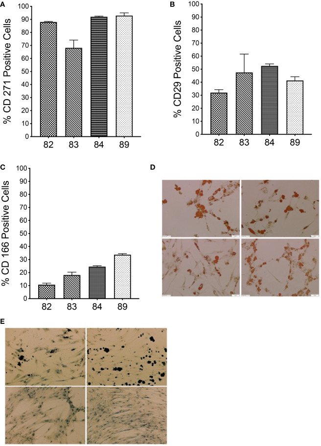
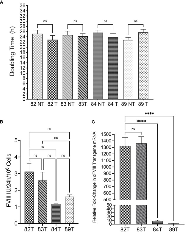
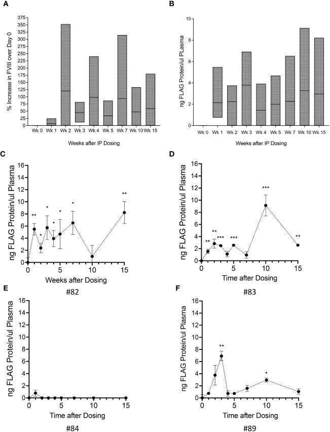
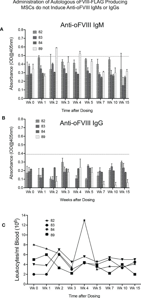
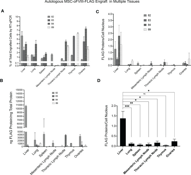
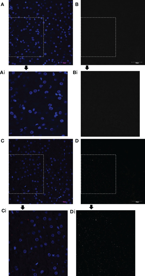
Results: We show that autologous BM-MSCs can be isolated, transduced with a lentivector to produce high levels of ovine (o)FVIII, extensively expanded, and transplanted into adult animals safely. The transplanted cells engraft in multiple organs, and they stably produce and secrete sufficient quantities of FVIII to yield elevated plasma FVIII levels for at least 15 weeks.

Discussion: These studies thus highlight the promise of cellular-based gene delivery approaches for treating HA.

Keywords: FVIII; Hemophilia A; bone marrow; cell therapy; efficacy & safety; gene therapy; mesenchymal stroma cell.


Conflict of interest statement

The authors declare that the research was conducted in the absence of any commercial or financial relationships that could be construed as a potential conflict of interest.

Figures

Figure 1
Figure 1
Characterization of autologous BM-derived MSCs. (A–C) Flow cytometric analysis of MSCs isolated from sheep bone marrow for CD271, CD29, and CD166 markers. (D) Oil Red O staining of MSCs from each sheep after differentiation into adipogenic lineage. (E) von Kossa staining of MSCs from each sheep after differentiation into osteocytic lineage. (n=4 biologic replicates; n=3 technical replicates).
Figure 2
Figure 2
Evaluation of Transduced Sheep Bone Marrow-Derived MSCs. (A) Transduction did not exert a statistically significant effect on cell doubling time (n=4 biologic replicates; n=4 technical replicates; p > 0.05). (B) FVIII activity in the supernatant of transduced cells was measured by one stage assay (aPTT) to determine the amount of oFVIII protein secreted by each MSCs population; levels of FVIII production differed markedly between the 4 individual sheep, but differences did not reach statistical significance (n=4 biologic replicates; n=3 technical replicates; p > 0.05). (C) RT-qPCR quantifying the expression of oFVIII mRNA showed a significant increase in the levels of oFVIII mRNA after transduction, with over a 1000-fold increase in the two MSCs lines that had higher levels of FVIII secretion, but more modest increases in the other two cell lines (n=4 biologic replicates; n=3 technical replicates; ****p < 0.0001). ns, not significant.
Figure 3
Figure 3
Administration of Autologous oFVIII-FLAG Producing MSCs Provide FVIII Protein in vivo. (A) Factor VIII activity measured in the plasma of all animals at weekly intervals after treatment with the autologous MSC-oFVIII-FLAG; the percent increase in FVIII activity over day 0 for each animal was calculated at each time point and the min to max increase in FVIII activity in the group of animals is shown (n=4 biological replicates). (B) Min to max levels oFVIII-FLAG protein in the plasma of animals after treatment measured by FLAG-specific ELISA (n=4 biological replicates, n=3 technical replicates). (C–F) Levels oFVIII-FLAG protein in the plasma of each individual animal measured by FLAG-specific ELISA (n=3) *p < 0.05, **p < 0.005, ***p < 0.0005 compared to week 0.
Figure 4
Figure 4
Administration of Autologous oFVIII-FLAG Producing MSCs do not Elicit an Immune Response or cause Noticeable Alterations in White Blood Cell Counts (A) Detection of anti-oFVIII IgMs in serum at the lowest dilution of 1:20, for each animal, at each timepoint, measured by ELISA (based on the background observed in a panel of negative control sheep, only signals above 0.49 were deemed to be positive) (n=3). (B) Detection of anti-oFVIII IgGs in serum (1:20 dilution) for each animal at each timepoint, measured by ELISA. Based on the background observed in a panel of negative control sheep, only signals above 0.3 were deemed to be positive (n=3). (C) White blood cell counts/ml of blood in each animal prior to and for 15 weeks after injections of autologous MSCs.
Figure 5
Figure 5
Quantification of Autologous MSC-oFVIII-FLAG Engraftment in Multiple Tissues by RT-qPCR, FLAG-specific ELISA, and Immunohistochemistry. (A) RT-qPCR was performed (n=3/organ/animal) using FLAG-tagged oFVIII specific primers on RNA isolated from different organs, and the percentage of MSC-oFVIII-FLAG engraftment was extrapolated using a standard curve prepared with RNA isolated from different percentages of MSC-oFVIII-FLAG mixed with sheep non-transduced MSCs. MSC-oFVIII-FLAG engrafted at detectable levels in each animal in every tissue examined. (B) Levels of oFVIII-FLAG protein in each tissue were determined by performing FLAG-specific ELISA on tissue homogenates, and the highest levels of oFVIII-FLAG protein were found in the liver of the treated animals (n=4 biologic replicates; n=3 technical replicates). (C) Immunohistochemistry with an antibody to the FLAG-tag protein was also performed in the various tissues to confirm the presence of oFVIII-FLAG in the different tissues; particle analysis in imageJ was used to quantify the total number of cells and the amount of FLAG protein to determine the quantity of FLAG protein per cell, and these analyses showed a pattern of engraftment similar to that found by ELISA. (D) Levels of oFVIII-FLAG protein were significantly higher in the liver of the recipients than any other tissue (n=4 biologic replicates; n=3 technical replicates; *p < 0.05, **p < 0.005, ***p < 0.0005). ns, not significant.
Figure 6
Figure 6
oFVIII-FLAG Immunohistochemistry Images of Liver Sections from a Control and a Treated Animal. (A) Representative images of the IHC staining of the liver from a control non-transplanted animal with an antibody to the FLAG-tag protein; (Ai) amplified inset area from A. (B, Bi) Same image as A and Ai but in black and white for better visualization after extracting the red channel and converting to grayscale in Photoshop. (C) Representative image of the IHC staining of the liver of treated animal with an antibody to the FLAG-tag protein (red); (Ci) amplified inset area from C. (D, Di) Same image as C and Ci but in black and white for better visualization after extracting the red channel and converting to grayscale in Photoshop. All images were acquired with an Olympus BX63 microscope Olympus with a 20x objective.

Similar articles

Cited by

References

    1. Giordano A, Galderisi U, Marino IR. From the laboratory bench to the patient’s bedside: An update on clinical trials with mesenchymal stem cells. J Cell Physiol (2007) 211(1):27–35. doi: 10.1002/jcp.20959 - DOI - PubMed
    1. Ringden O, Moll G, Gustafsson B, Sadeghi B. Mesenchymal stromal cells for enhancing hematopoietic engraftment and treatment of graft-Versus-Host disease, hemorrhages and acute respiratory distress syndrome. Front Immunol (2022) 13:839844. doi: 10.3389/fimmu.2022.839844 - DOI - PMC - PubMed
    1. Almeida-Porada G, Atala AJ, Porada CD. Therapeutic mesenchymal stromal cells for immunotherapy and for gene and drug delivery. Mol Ther Methods Clin Dev (2020) 16:204–24. doi: 10.1016/j.omtm.2020.01.005 - DOI - PMC - PubMed
    1. Di Nicola M, Carlo-Stella C, Magni M, Milanesi M, Longoni PD, Matteucci P, et al. . Human bone marrow stromal cells suppress T-lymphocyte proliferation induced by cellular or nonspecific mitogenic stimuli. Blood (2002) 99(10):3838–43. doi: 10.1182/blood.V99.10.3838 - DOI - PubMed
    1. Joswig A-J, Mitchell A, Cummings KJ, Levine GJ, Gregory CA, Smith R, et al. . Repeated intra-articular injection of allogeneic mesenchymal stem cells causes an adverse response compared to autologous cells in the equine model. Stem Cell Res Ther (2017) 8(1):42. doi: 10.1186/s13287-017-0503-8 - DOI - PMC - PubMed

Publication types