Review
. 2022;2(1):96.
doi: 10.1038/s43586-022-00175-x. Epub 2022 Dec 8.

Liquid chromatography-tandem mass spectrometry for clinical diagnostics

Affiliations
Review

Liquid chromatography-tandem mass spectrometry for clinical diagnostics

Stefani N Thomas et al. Nat Rev Methods Primers. 2022.

Abstract

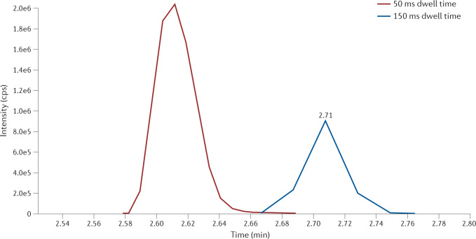
Mass spectrometry is a powerful analytical tool used for the analysis of a wide range of substances and matrices; it is increasingly utilized for clinical applications in laboratory medicine. This Primer includes an overview of basic mass spectrometry concepts, focusing primarily on tandem mass spectrometry. We discuss experimental considerations and quality management, and provide an overview of some key applications in the clinic. Lastly, the Primer discusses significant challenges for implementation of mass spectrometry in clinical laboratories and provides an outlook of where there are emerging clinical applications for this technology.

Keywords: Mass spectrometry; Medical research.


Conflict of interest statement

Competing interestsW.A.C. declares consulting for and research support from Thermo Fisher Scientific, consulting for Roche Diagnostics, and research support from Shimadzu Scientific Instruments. The other authors declare no competing interests.

Figures

Fig. 1
Fig. 1. Depiction of a liquid chromatography–tandem mass spectrometry system.
Electrospray ionization shown as the ion source with a triple quadrupole analyser.
Fig. 2
Fig. 2. Overview of mass analysis.
a, A triple quadrupole mass spectrometer and how each quadrupole transmits ions. b, Selected reaction monitoring using testosterone as an example. m/z, mass to charge ratio.
Fig. 3
Fig. 3. Effect of the dwell time being appropriate for the width of the peak versus too long.
The peak acquired with 50 ms dwell time provides a sufficient point across the peak. A dwell time of 150 ms results in a poorly defined peak with only five points.
Fig. 4
Fig. 4. Quantification of testosterone by liquid chromatography–tandem mass spectrometry.
a, Chromatogram of testosterone. b, Signal-to-noise ratio for determination. c, Calculation of testosterone concentration in patient sample based on calibration curve. Peak area ratio = analyte peak area divided by internal standard peak area.
Fig. 5
Fig. 5. Analysis of leucine isomers.
Example liquid chromatography–tandem mass spectrometry data generated from the analysis of leucine isomers with counts on the y axis (abundance of response) and time on the x axis. a, Dark blue line: transition 132.1 (quadrupole 1) to 44.1 (quadrupole 3) for equimolar concentrations of alloisoleucine (0.16 min), isoleucine (0.25 min) and leucine (0.40 min). Light blue line: transition 132.1 (quadrupole 1) to 43.1 (quadrupole 3), which is used as a qualifying transition for leucine only. The differences in recorded abundance are caused by the ionization and dissociation differences at the applied collision energy for the selected transitions. b, Transition 142.1 (quadrupole 1) to 78.1 (quadrupole 3) for the 10-deuteron labelled (‘D10’) alloisoleucine internal standard (0.14 min), which is used to normalize the recovery of alloisoleucine. c, Transition 142.1 (quadrupole 1) to 96.1 (quadrupole 3) for both D10-alloisoleucine (0.14 min) and D10-leucine (0.37 min); this transition acts as a normalizer for leucine quantification and as a qualifying transition for the alloisoleucine internal standard. The area under the curve for each peak is used for quantification calculations. MRM, multiple reaction monitoring; XIC, extracted ion chromatogram.

References

    1. Haag AM. Mass analyzers and mass spectrometers. Adv. Exp. Med. Biol. 2016;919:157–169. doi: 10.1007/978-3-319-41448-5_7. - DOI - PubMed
    1. Chardwaj C, Hanley L. Ion sources for mass spectrometric identification and imaging of molecular species. Nat. Prod. Rep. 2014;31:756–767. doi: 10.1039/C3NP70094A. - DOI - PubMed
    1. Hage, D.S. in Contemporary Practice in Clinical Chemistry 4th edn Ch. 8 (eds Marzinke, M. A. & Clarke, W.) 135–157 (Elsevier, 2020).
    1. CLSI. Liquid Chromatography–Mass Spectrometry Methods; Approved Guideline CLSI Document C62-A (Clinical and Laboratory Standards Institute, 2014).
    1. Lynch KL. Accreditation and quality assurance for clinical liquid chromatography–mass spectrometry laboratories. Clin. Lab. Med. 2018;38:515–526. doi: 10.1016/j.cll.2018.05.002. - DOI - PubMed