Endothelial β-arrestins regulate mechanotransduction by the type II bone morphogenetic protein receptor in primary cilia
- PMID: 36532314
- PMCID: PMC9751664
- DOI: 10.1002/pul2.12167
Endothelial β-arrestins regulate mechanotransduction by the type II bone morphogenetic protein receptor in primary cilia
Abstract
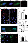
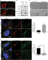
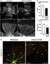
Modulation of endothelial cell behavior and phenotype by hemodynamic forces involves many signaling components, including cell surface receptors, intracellular signaling intermediaries, transcription factors, and epigenetic elements. Many of the signaling mechanisms that underlie mechanotransduction by endothelial cells are inadequately defined. Here we sought to better understand how β-arrestins, intracellular proteins that regulate agonist-mediated desensitization and integration of signaling by transmembrane receptors, may be involved in the endothelial cell response to shear stress. We performed both in vitro studies with primary endothelial cells subjected to β-arrestin knockdown, and in vivo studies using mice with endothelial specific deletion of β-arrestin 1 and β-arrestin 2. We found that β-arrestins are localized to primary cilia in endothelial cells, which are present in subpopulations of endothelial cells in relatively low shear states. Recruitment of β-arrestins to cilia involved its interaction with IFT81, a component of the flagellar transport protein complex in the cilia. β-arrestin knockdown led to marked reduction in shear stress response, including induction of NOS3 expression. Within the cilia, β-arrestins were found to associate with the type II bone morphogenetic protein receptor (BMPR-II), whose disruption similarly led to an impaired endothelial shear response. β-arrestins also regulated Smad transcription factor phosphorylation by BMPR-II. Mice with endothelial specific deletion of β-arrestin 1 and β-arrestin 2 were found to have impaired retinal angiogenesis. In conclusion, we have identified a novel role for endothelial β-arrestins as key transducers of ciliary mechanotransduction that play a central role in shear signaling by BMPR-II and contribute to vascular development.
Keywords: BMPR2; beta‐arrestin; endothelial cells; primary cilia; shear stress.
© 2022 The Authors. Pulmonary Circulation published by Wiley Periodicals LLC on behalf of the Pulmonary Vascular Research Institute.
Conflict of interest statement
The authors declare no conflict of interest.
Figures


Similar articles
-
Recruitment of β-Arrestin into Neuronal Cilia Modulates Somatostatin Receptor Subtype 3 Ciliary Localization.Mol Cell Biol. 2015 Oct 26;36(1):223-35. doi: 10.1128/MCB.00765-15. Print 2016 Jan 1. Mol Cell Biol. 2015. PMID: 26503786 Free PMC article.
-
β-arrestin is critical for early shear stress-induced Akt/eNOS activation in human vascular endothelial cells.Biochem Biophys Res Commun. 2017 Jan 29;483(1):75-81. doi: 10.1016/j.bbrc.2017.01.003. Epub 2017 Jan 4. Biochem Biophys Res Commun. 2017. PMID: 28062183
-
Distinct and shared roles of β-arrestin-1 and β-arrestin-2 on the regulation of C3a receptor signaling in human mast cells.PLoS One. 2011 May 12;6(5):e19585. doi: 10.1371/journal.pone.0019585. PLoS One. 2011. PMID: 21589858 Free PMC article.
-
Beta-arrestin signaling and regulation of transcription.J Cell Sci. 2007 Jan 15;120(Pt 2):213-8. doi: 10.1242/jcs.03338. J Cell Sci. 2007. PMID: 17215450 Review.
-
Beta-Arrestins and Receptor Signaling in the Vascular Endothelium.Biomolecules. 2020 Dec 23;11(1):9. doi: 10.3390/biom11010009. Biomolecules. 2020. PMID: 33374806 Free PMC article. Review.
Cited by
-
Impact of Hedgehog modulators on signaling pathways in primary murine and human hepatocytes in vitro: insights into liver metabolism.Arch Toxicol. 2025 Mar;99(3):1105-1116. doi: 10.1007/s00204-024-03931-y. Epub 2024 Dec 23. Arch Toxicol. 2025. PMID: 39714734 Free PMC article.
-
TNF-alpha promotes cilia elongation via mixed lineage kinases signaling in mouse fibroblasts and human RPE-1 cells.Cytoskeleton (Hoboken). 2024 Nov;81(11):639-647. doi: 10.1002/cm.21873. Epub 2024 May 20. Cytoskeleton (Hoboken). 2024. PMID: 38767050
-
β-arrestin2: an emerging player and potential therapeutic target in inflammatory immune diseases.Acta Pharmacol Sin. 2025 Sep;46(9):2347-2362. doi: 10.1038/s41401-024-01390-w. Epub 2024 Sep 30. Acta Pharmacol Sin. 2025. PMID: 39349766 Review.
-
ARL13B controls male reproductive tract physiology through primary and Motile Cilia.Commun Biol. 2024 Oct 14;7(1):1318. doi: 10.1038/s42003-024-07030-7. Commun Biol. 2024. PMID: 39397107 Free PMC article.
References
-
- Ando J, Yamamoto K. Flow detection and calcium signalling in vascular endothelial cells. Cardiovasc Res. 2013;99(2):260–8. - PubMed
-
- Langille BL, Adamson SL. Relationship between blood‐flow direction and endothelial‐cell orientation at arterial branch sites in rabbits and mice. Circ Res. 1981;48(4):481–8. - PubMed
-
- Levesque MJ, Nerem RM. The elongation and orientation of cultured endothelial cells in response to shear stress. J Biomech Eng. 1985;107(4):341–7. - PubMed
Grants and funding
LinkOut - more resources
Full Text Sources
Miscellaneous

