

Investigations on the substrate binding sites of hemolysin B, an ABC transporter, of a type 1 secretion system
- PMID: 36532430
- PMCID: PMC9751043
- DOI: 10.3389/fmicb.2022.1055032
Investigations on the substrate binding sites of hemolysin B, an ABC transporter, of a type 1 secretion system
Abstract
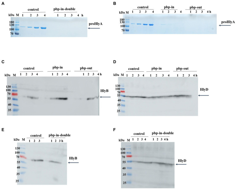
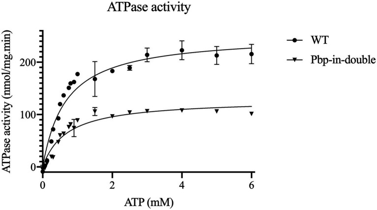
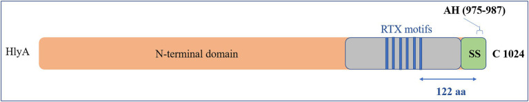
The ABC transporter hemolysin B (HlyB) is the key protein of the HlyA secretion system, a paradigm of type 1 secretion systems (T1SS). T1SS catalyze the one-step substrate transport across both membranes of Gram-negative bacteria. The HlyA T1SS is composed of the ABC transporter (HlyB), the membrane fusion protein (HlyD), and the outer membrane protein TolC. HlyA is a member of the RTX (repeats in toxins) family harboring GG repeats that bind Ca2+ in the C-terminus upstream of the secretion signal. Beside the GG repeats, the presence of an amphipathic helix (AH) in the C-terminus of HlyA is essential for secretion. Here, we propose that a consensus length between the GG repeats and the AH affects the secretion efficiency of the heterologous RTX secreted by the HlyA T1SS. Our in silico studies along with mutagenesis and biochemical analysis demonstrate that there are two binding pockets in the nucleotide binding domain of HlyB for HlyA. The distances between the domains of HlyB implied to interact with HlyA indicated that simultaneous binding of the substrate to both cytosolic domains of HlyB, the NBD and CLD, is possible and required for efficient substrate secretion.
Keywords: ABC transporter; bacterial secretion systems; protein secretion; putative binding pockets; substrate interaction.
Copyright © 2022 Pourhassan N, Hachani, Spitz, Smits and Schmitt.
Conflict of interest statement
The authors declare that the research was conducted in the absence of any commercial or financial relationships that could be construed as a potential conflict of interest.
Figures


Similar articles
-
Type 1 secretion necessitates a tight interplay between all domains of the ABC transporter.Sci Rep. 2024 Apr 18;14(1):8994. doi: 10.1038/s41598-024-59759-0. Sci Rep. 2024. PMID: 38637678 Free PMC article.
-
Quantification and Surface Localization of the Hemolysin A Type I Secretion System at the Endogenous Level and under Conditions of Overexpression.Appl Environ Microbiol. 2022 Feb 8;88(3):e0189621. doi: 10.1128/AEM.01896-21. Epub 2021 Dec 1. Appl Environ Microbiol. 2022. PMID: 34851699 Free PMC article.
-
Interdomain regulation of the ATPase activity of the ABC transporter haemolysin B from Escherichia coli.Biochem J. 2016 Aug 15;473(16):2471-83. doi: 10.1042/BCJ20160154. Epub 2016 Jun 8. Biochem J. 2016. PMID: 27279651
-
Type 1 protein secretion in bacteria, the ABC-transporter dependent pathway (review).Mol Membr Biol. 2005 Jan-Apr;22(1-2):29-39. doi: 10.1080/09687860500042013. Mol Membr Biol. 2005. PMID: 16092522 Review.
-
A molecular understanding of the catalytic cycle of the nucleotide-binding domain of the ABC transporter HlyB.Biochem Soc Trans. 2005 Nov;33(Pt 5):990-5. doi: 10.1042/BST20050990. Biochem Soc Trans. 2005. PMID: 16246029 Review.
Cited by
-
Progress toward a vaccine for extraintestinal pathogenic E. coli (ExPEC) II: efficacy of a toxin-autotransporter dual antigen approach.Infect Immun. 2024 May 7;92(5):e0044023. doi: 10.1128/iai.00440-23. Epub 2024 Apr 9. Infect Immun. 2024. PMID: 38591882 Free PMC article.
-
Type 1 secretion necessitates a tight interplay between all domains of the ABC transporter.Sci Rep. 2024 Apr 18;14(1):8994. doi: 10.1038/s41598-024-59759-0. Sci Rep. 2024. PMID: 38637678 Free PMC article.
-
A step forward to the optimized HlyA type 1 secretion system through directed evolution.Appl Microbiol Biotechnol. 2023 Aug;107(16):5131-5143. doi: 10.1007/s00253-023-12653-7. Epub 2023 Jul 5. Appl Microbiol Biotechnol. 2023. PMID: 37405436 Free PMC article.
References
-
- Abràmoff M. D., Magalhães P. J., Ram S. J. (2004). Image processing with ImageJ. Biophoton. Int. 11, 36–42.
-
- Benabdelhak H., Kiontke S., Horn C., Ernst R., Blight M. A., Holland I. B., et al. (2003). A specific interaction between the NBD of the ABC-transporter HlyB and a C-terminal fragment of its transport substrate haemolysin a. J. Mol. Biol. 327, 1169–1179. doi: 10.1016/S0022-2836(03)00204-3, PMID: - DOI - PubMed
LinkOut - more resources
Full Text Sources
Miscellaneous