

Enhanced Intraoperative Communication of Tumor Margins Using 3D Scanning and Mapping: The Computer-Aided Design Margin
- PMID: 36533659
- PMCID: PMC10277314
- DOI: 10.1002/lary.30511
Enhanced Intraoperative Communication of Tumor Margins Using 3D Scanning and Mapping: The Computer-Aided Design Margin
Abstract
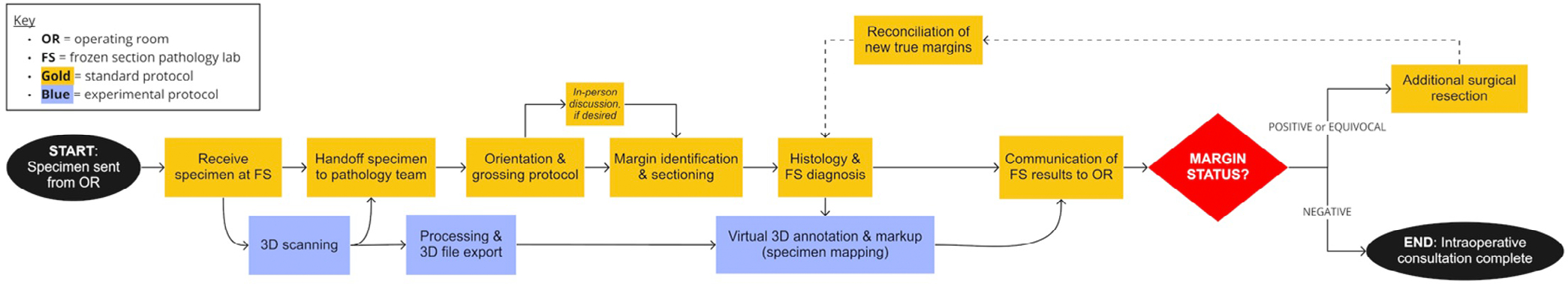
We have implemented a novel intraoperative communication system for head and neck surgical pathology using 3D scanning technology and computer-aided design (CAD) software. Here, we highlight our CAD margin protocol, in which we 3D scan head and neck oncologic surgical specimens and map frozen section results. This enhances the communication of margin status between surgeons and pathologists and delivers visual anatomic guidance for re-resection when needed. Laryngoscope, 133:1914-1918, 2023.
Keywords: 3D scanning; 3D specimen mapping; communication; frozen section; head and neck; margin analysis.
© 2022 The American Laryngological, Rhinological and Otological Society, Inc.
Figures


References
-
- Looser KG, Shah JP, Strong EW. The significance of “positive” margins in surgically resected epidermoid carcinomas. Head Neck Surg. 1978;1(2):107–111. - PubMed
-
- Amit M, Na’ara S, Leider-Trejo L, et al. Improving the rate of negative margins after surgery for oral cavity squamous cell carcinoma: a prospective randomized controlled study. Head Neck. 2016;38(S1):E1803–E1809. - PubMed
-
- Kerawala CJ, Ong TK. Relocating the site of frozen sections—is there room for improvement? Head Neck. 2001;23(3):230–232. - PubMed
-
- Buchakjian MR, Tasche KK, Robinson RA, Pagedar NA, Sperry SM. Association of main specimen and tumor bed margin status with local recurrence and survival in oral cancer surgery. JAMA Otolaryngol Head Neck Surg. 2016;142(12):1191–1198. - PubMed
Publication types
MeSH terms
Grants and funding
LinkOut - more resources
Full Text Sources
Medical
Miscellaneous
