. 2023 Jan 3;62(1):134-144.
doi: 10.1021/acs.biochem.2c00633. Epub 2022 Dec 19.

Bifunctional Epimerase/Reductase Enzymes Facilitate the Modulation of 6-Deoxy-Heptoses Found in the Capsular Polysaccharides of Campylobacter jejuni

Affiliations

Bifunctional Epimerase/Reductase Enzymes Facilitate the Modulation of 6-Deoxy-Heptoses Found in the Capsular Polysaccharides of Campylobacter jejuni

Dao Feng Xiang et al. Biochemistry. .

Abstract

Campylobacter jejuni is a human pathogen and the leading cause of food poisoning in the United States and Europe. Surrounding the exterior surface of this bacterium is a capsular polysaccharide (CPS) that consists of a repeating sequence of common and unusual carbohydrate segments. At least 10 different heptose sugars have thus far been identified in the various strains of C. jejuni. The accepted biosynthetic pathway for the construction of the 6-deoxy-heptoses begins with the 4,6-dehydration of GDP-d-glycero-d-manno-heptose by a dehydratase, followed by an epimerase that racemizes C3 and/or C5 of the product GDP-6-deoxy-4-keto-d-lyxo-heptose. In the final step, a C4-reductase catalyzes the NADPH reduction of the resulting 4-keto product. However, in some strains and serotypes of C. jejuni, there are two separate C4-reductases with different product specificities in the gene cluster for CPS formation. Five pairs of these tandem C4-reductases were isolated, and the catalytic properties were ascertained. In four out of five cases, one of the two C4-reductases is able to catalyze the isomerization of C3 and C5 of GDP-6-deoxy-4-keto-d-lyxo-heptose, in addition to the catalysis of the reduction of C4, thus bypassing the requirement for a separate C3/C5-isomerase. In each case, the 3'-end of the gene for the first C4-reductase contains a poly-G tract of 8-10 guanine residues that may be used to control the expression and/or catalytic activity of either C4-reductase. The three-dimensional structure of the C4-reductase from serotype HS:15, which only does a reduction of C4, was determined to 1.45 Å resolution in the presence of NADPH and GDP.


Conflict of interest statement

The authors declare no competing conflicts of interest.

Figures

Figure 1:
Figure 1:
Structures of the repeating capsular polysaccharides in the HS:4 and HS:15 serotypes. The HS:4 serotype contains N-acetyl-d-glucosamine and 6-deoxy-d-ido-heptose (or l-glycero-d-ido-heptose), whereas the HS:15 serotype contains l-arabinose and 6-deoxy-l-gulo-heptose (12, 13). The CPS from serotype HS:4 has also been shown to be decorated with an O-methyl phosphoramidate moiety on the ido-heptose at C2 or C7 (12).
Figure 2:
Figure 2:
Biosynthetic pathway for formation of 6-deoxy-heptoses.
Figure 3:
Figure 3:
The structures of six GDP-6-deoxy-heptoses that have been isolated from the catalytic activity of eight C4-reductases from various strains of C. jejuni (6, 16).
Figure 4:
Figure 4:
Sequence similarity network for 25 C4-reductases identified within various strains of C. jejuni at a sequence identity cutoff of 89%. The green colored nodes denote C4-reductases that have previously been isolated and the products characterized (11, 16). The yellow-colored nodes indicate C4-reductases that are characterized in this investigation. The grey-colored nodes are for reductases likely involved in the biosynthesis of 3,6-dideoxy heptoses and those colored blue are additional C4-reductases that have not been purified. The product specificities for the numbered clusters are as follows: Group-1 (GDP-6-deoxy-d-altro-heptose (4)); Group-2 (GDP-6-deoxy-l-galacto-heptose (6)); Group-3 (GDP-6-deoxy-d-ido-heptose (5)); Group-4 (GDP-6-deoxy-l-gulo-heptose (8)); Group-5 (GDP-6-deoxy-d-manno-heptose (3)); Group-6 (GDP-6-deoxy-l-gluco-heptose (7)); and Group-7 (GDP-6-deoxy-d-ido-heptose (5)).
Figure 5:
Figure 5:
Demonstration of the ability of the HS:10B C4-reductase to catalyze the isomerization of C3 and C5 of GDP-6-deoxy-4-keto-d-lyxo-heptose (2). The enzyme was incubated with compound 2 in the presence of NADP+ at pH 7.5. (A) Control experiment showing a portion of the 1H NMR spectrum of compound 2 prior to the addition of enzyme. The doublet at ~5.48 ppm is from the hydrogen attached to C1. (B) A portion of the 1H NMR spectrum of compound 2 after the addition of 4 μM HS:10B C4-reductase in the presence of NADP+. The unresolved doublet of doublets at ~5.38 ppm is for the C3-isomerized product and the triplet at ~5.31 ppm is for the C5-isomerized product. The triplet at ~5.02 ppm is from the C3/C5-isomerized product (8). Additional details are provided in the text.
Figure 6:
Figure 6:
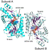
Ribbon representation of the C4-reductase from serotype HS:15. The enzyme functions as a dimer with C2 symmetry. In the view shown, the two-fold rotational axis is perpendicular to the plane of the page. The ligands are depicted in space filling representations.
Figure 7:
Figure 7:
Superposition of the ribbon drawings for the C4-reductase and the GDP-l-fucose synthase. The α-carbon traces for the reductase and fucose synthase and their bound ligands are highlighted in teal and gray, respectively.
Figure 8:
Figure 8:
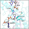
Closeup view of the active site. Tyr137 and Lys141 are found, with only a few exception, in all SDR superfamily members. Cys110 and His180 are typically found in those C4-reductases that catalyze epimerizations. Note that the nicotinamide ring of the NADPH adopts the syn conformation, which is another characteristic for members of the SDR superfamily.
Figure 9.
Figure 9.
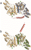
Structural alignment of the C4-reductase Cj1428 from C. jejuni serotype HS:2 and Alphafold2 structural predictions of the HS:29A and HS:8B C4-reductases. (A) The structure of Cj1428 (PDB id: 7M13) is depicted as a dimer colored in wheat with bound ligands shown as green sticks. The predicted structure of the HS:29A C4-reductase monomer is shown in blue (residues 1–341) with the extra amino acids at the C-terminus of the protein shown in red (residues 342–378). (B) C4-reductase Cj1428 from serotype HS:2 as a dimer in wheat (PDB id: 7M13) with bound ligands shown as green sticks. Shown in the green ribbon diagram is the Alphafold2 structural prediction for the HS:8B C4-reductase (residues 29–374). The amino acids at the N-terminus are colored red (residues 1–28).

References

    1. Heimesaat MM; Backert S; Alter T; Bereswill S Human Campylobacteriosis-A Serious Infectious Threat in a One Health Perspective. Curr. Top. Microbiol. Immunol 2021, 431, 1–23. - PubMed
    1. Burnham PM; Hendrixson DR Campylobacter jejuni: Collective Components Promoting a Successful Enteric Lifestyle. Nat. Rev. Microbiol 2018, 16, 551–565. - PubMed
    1. Monteiro MA; Noll A; Laird RM; Pequegnat B; Ma ZC; Bertolo L; DePass C; Omari E; Gabryelski P; Redkyna O; Jiao YN; Borrelli S; Poly F; Guerry P Campylobacter jejuni Capsule Polysaccharide Conjugate Vaccine. In Carbohydrate-based Vaccines: from Concept to Clinic; American Chemical Society: Washington, DC, 2018, pp 249–271.
    1. Michael F; Szymanski CM; Li JJ; Chan KH; Khieu NH; Larocque S; Wakarchuk WW; Brisson JR; Monteiro MA The Structures of the Lipooligosaccharide and Capsule Colysaccharide of Campylobacter jejuni Genome Sequenced Strain NCTC 11168. Eur. J. Biochem 2002, 269, 5119–5136. - PubMed
    1. Aspinall GO; Monteiro MA; Pang H Lipo-oligosaccharide of the Campylobacter lari type strain ATCC 35221. Structure of the liberated oligosaccharide and an associated extracellular polysaccharide. Carbohydrate Research 1995, 279, 245–264. - PubMed

Publication types