Review
. 2022 Oct 1;12(4):e2022162.
doi: 10.5826/dpc.1204a162. eCollection 2022 Nov.

Challenges for New Adopters in Pre-Surgical Margin Assessment by Handheld Reflectance Confocal Microscope of Basal Cell Carcinoma; A Prospective Single-center Study

Affiliations
Review

Challenges for New Adopters in Pre-Surgical Margin Assessment by Handheld Reflectance Confocal Microscope of Basal Cell Carcinoma; A Prospective Single-center Study

Nina Anika Richarz et al. Dermatol Pract Concept. .

Abstract

Introduction: In vivo reflectance confocal microscopy (RCM) is a useful tool for assessing pre-surgical skin tumor margins when performed by a skilled, experienced user. The technique, however, poses significant challenges to novice users, particularly when a handheld RCM (HRCM) device is used.

Objectives: To evaluate the performance of an HRCM device operated by a novice user to delineate basal cell carcinoma (BCC) margins before Mohs micrographic surgery (MMS).

Methods: Prospective study of 17 consecutive patients with a BCC in a high-risk facial area (the H zone) in whom tumor margins were assessed by HRCM and dermoscopy before MMS. Predicted surgical defect areas (cm2) were calculated using standardized photographic digital documentation and compared to final defect areas after staged excision.

Results: No significant differences were observed between median HRCM-predicted and observed surgical defect areas (2.95 cm2 [range: 0.83-17.52] versus 2.52 cm2 [range 0.71-14.42]; P = 0.586). Dermoscopy, by contrast, produced significantly underestimated values (median area of 1.34 cm2 [0.41-4.64] versus 2.52 cm2 [range 0.71-14.42]; P < 0.001). Confounders leading to poor agreement between predicted and observed areas were previous treatment (N = 5), a purely infiltrative subtype (N = 1), and abundant sebaceous hyperplasia (N = 1).

Conclusions: Even in the hands of a novice user, HRCM is more accurate than dermoscopy for delineating lateral BCCs margins in high-risk areas and performs well at predicting final surgical defects.

Keywords: basal cell carcinoma; margin control; micrographic Mohs surgery; reflectance confocal microscopy.


Conflict of interest statement

Competing interests: None.

Figures
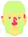
Figure 1
Figure 1
Locations of basal cell carcinomas (red circles) included in the study. High-risk areas are shown in orange.
Figure 2
Figure 2
Representative cases of basal cell carcinoma (BCC) showing good agreement between surgical defect areas predicted by handheld reflectance confocal microscopy (HRCM) and the defect areas after Mohs micrographic surgery (MMS) (A–D), overestimated defect areas (E–H), and underestimated defect areas (I–L). (A) Similarity between the HRCM-predicted surgical defect area (blue line) and the final area (B) in a BCC with evident tumor islands seen by RCM (C) and a nodular subtype identified in the intraoperative frozen section (D). (E) HRCM-predicted surgical defect area in a BCC previously treated with radiotherapy that was significantly larger than the final defect area after MMS (F). HRCM images of radiation-induced dermal fibrosis (G) were mistaken for collagen surrounding deep tumor islands, but these were ruled out by histology (H). The HRCM–predicted surgical defect area in (I) was significantly smaller than the final area (J) in a superficial, infiltrative BCC visible by HRCM (K) and histology (L).

References

    1. Lomas A, Leonardi-Bee J, Bath-Hextall F. A systematic review of worldwide incidence of nonmelanoma skin cancer. Br J Dermatol. 2012;166(5):1069–1080. doi: 10.1111/j.1365-2133.2012.10830.x. - DOI - PubMed
    1. Van Loo E, Mosterd K, Krekels GAM, et al. Surgical excision versus Mohs’ micrographic surgery for basal cell carcinoma of the face: A randomised clinical trial with 10 year follow-up. Eur J Cancer. 2014;50(17):3011–3020. doi: 10.1016/j.ejca.2014.08.018. - DOI - PubMed
    1. Pan ZY, Lin JR, Cheng TT, Wu JQ, Wu WY. In vivo reflectance confocal microscopy of basal cell carcinoma: Feasibility of preoperative mapping of cancer margins. Dermatologic Surg. 2012;38(12):1945–1950. doi: 10.1111/j.1524-4725.2012.02587.x. - DOI - PMC - PubMed
    1. Iftimia N, Yélamos O, Chen C-SJ, et al. Handheld optical coherence tomography–reflectance confocal microscopy probe for detection of basal cell carcinoma and delineation of margins. J Biomed Opt. 2017;22(7):076006. doi: 10.1117/1.JBO.22.7.076006. - DOI - PMC - PubMed
    1. Venturini M, Gualdi G, Zanca A, Lorenzi L, Pellacani G, Calzavara-Pinton PG. A new approach for presurgical margin assessment by reflectance confocal microscopy of basal cell carcinoma. Br J Dermatol. 2016;174(2):380–385. doi: 10.1111/bjd.14244. - DOI - PubMed

LinkOut - more resources