. 2022 Oct 1;12(4):e2022203.
doi: 10.5826/dpc.1204a203. eCollection 2022 Nov.

Dermoscopy of the Diverse Spectrum of Cutaneous Tuberculosis in the Skin of Color

Affiliations

Dermoscopy of the Diverse Spectrum of Cutaneous Tuberculosis in the Skin of Color

Rashmi Jindal et al. Dermatol Pract Concept. .

Abstract

Introduction: Cutaneous tuberculosis is an uncommon form of tuberculosis, accounting for 1%-2% of all forms of extra-pulmonary tuberculosis. Knowledge of the dermoscopic characteristics of different clinical types of cutaneous tuberculosis can help timely diagnosis resulting in better outcomes.

Objectives: To characterize the Dermoscopy findings in different clinical types of cutaneous tuberculosis in dark skin phototypes.

Methods: All clinically suspected and biopsy confirmed cases of cutaneous tuberculosis seen from July 2019 through December 2021 were retrospectively recruited. Information including age, gender, disease duration, site and morphology of lesions, and presence of concomitant tuberculosis elsewhere was noted. Two investigators retrospectively reviewed the dermoscopic characteristics of these cases.

Results: Twenty-two patients comprised of 12 women and 10 men met the inclusion criteria. Lupus vulgaris was the commonest presentation of cutaneous tuberculosis seen in 13 patients. Five had scrofuloderma, 2 had tuberculosis verrucosa cutis and 1 patient each had lichen scrofulosorum and papulo-necrotic tuberculid. Yellow-orange structureless areas (100%), linear/dot vessels (100%), white scales (92.3%), and white structureless areas (84.6%) were the predominant dermoscopy findings in lupus vulgaris. In scrofuloderma, linear vessels and white structureless areas were visible in all cases. Dirty white scales with a papillated surface were characteristically seen in tuberculosis verrucosa cutis, with 1 of the 2 patients each showing vessels and yellow-orange structureless areas. White globules with surrounding erythema were seen in lichen scrofulosorum and yellow-orange structureless areas with keratin plugs in papulo-necrotic tuberculid.

Conclusions: A thorough understanding of the characteristic dermoscopy of cutaneous tuberculosis can help suspect the diagnosis early resulting in better management opportunity.

Keywords: cutaneous tuberculosis; dermoscopy; lupus vulgaris; scrofuloderma.


Conflict of interest statement

Competing interests: None.

Figures

Figure 1
Figure 1
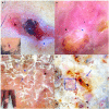
(A–D) Dermoscopy of lupus vulgaris showing yellow-orange structureless areas (black star), linear vessels with and without branches (black arrow), white structureless areas (blue star) and lines (blue arrowheads), follicular plugging (black circle), white scales (blue arrow), dot vessels (black square) and peri-lesional halo (blue rectangle). (A, inset) A well-defined plaque with atrophy and an advancing erythematous margin
Figure 2
Figure 2
Dermoscopy of scrofuloderma showing sero-sanguineous crust representing the sinus with yellow-orange structureless areas (black star), white structureless areas (blue star) and linear vessels (black arrow) (A,B), clinical image showing a central sinus surrounded by erythema and scarring (A, inset). Dermoscopy of tuberculosis verrucosa cutis showing thick dirty white scales (blue arrow), yellow orange structureless areas (black star), white structureless areas (blue star) and dot/curved vessels (blue square) (C,D), clinical image showing hyperkeratotic plaque over middle finger (C, inset).
Figure 3
Figure 3
(A) Dermoscopy of lichen scrofulosorum with monomorphic white clods, some having a central black dot (black circle) and some peripheral erythema (black square) and increased surrounding pigment network (black star), inset showing grouped papules over trunk. (B,C) Dermoscopy of papulo-necrotic tuberculid showing yellow-orange structureless areas (black star), inset showing purpuric macules over soles and keratin plugs (black circle) with inset showing hyperkeratotic papules.

Similar articles

Cited by

References

    1. Brito AC, Oliveira CMM, Unger DA, Bittencourt MJS. Cutaneous tuberculosis: epidemiological, clinical, diagnostic and therapeutic update. An Bras Dermatol. 2022;97(2):129–144. doi: 10.1016/j.abd.2021.07.004. - DOI - PMC - PubMed
    1. Supekar BB, Wankhade VH, Singh RP, Ghanate TD, Bhat D. Clinical spectrum of cutaneous tuberculosis in Central India: A retrospective study. Indian Dermatol Online J. 2021;12(6):826–833. doi: 10.4103/2229-5178.330811. - DOI - PMC - PubMed
    1. Errichetti E, Ankad BS, Jha AK, et al. International Dermoscopy Society criteria for non-neoplastic dermatoses (general dermatology): validation for skin of color through a Delphi expert consensus. Int J Dermatol. 2022;61(4):461–471. doi: 10.1111/ijd.15729. - DOI - PubMed
    1. Ankad BS, Adya KA, Gaikwad SS, Inamadar AC, Manjula R. Lupus Vulgaris in Darker Skin: Dermoscopic and Histopathologic Incongruity. Indian Dermatol Online J. 2020;11(6):948–952. doi: 10.4103/idoj.IDOJ_100_20. - DOI - PMC - PubMed
    1. Errichetti E, Stinco G. Dermatoscopy of Granulomatous Disorders. Dermatol Clin. 2018;36(4):369–375. doi: 10.1016/j.det.2018.05.004. - DOI - PubMed