

Dermoscopy and Reflectance Confocal Microscopy to Estimate Breslow Index and Mitotic Rate in Primary Melanoma
- PMID: 36534562
- PMCID: PMC9681198
- DOI: 10.5826/dpc.1204a174
Dermoscopy and Reflectance Confocal Microscopy to Estimate Breslow Index and Mitotic Rate in Primary Melanoma
Abstract
Introduction: Non-invasive imaging techniques offer the possibility to optimize the first approach to melanoma. Reflectance Confocal Microscopy (RCM) has a promising role in predicting the main prognostic events in the dermo-epidermal and papillary dermis.
Objectives: To identify pre-surgical criteria that can predict the main prognostic features of melanoma.
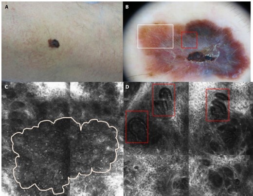
Methods: A retrospective cohort-study evaluated dermoscopic, confocal and histopathological characteristics of consecutively diagnosed sporadic melanomas. RCM-melanoma patterns classified into 1) dendritic-cell, 2) round-cell, 3) dermal nest and 4) combined type. Acral, facial and mucosal locations were excluded.
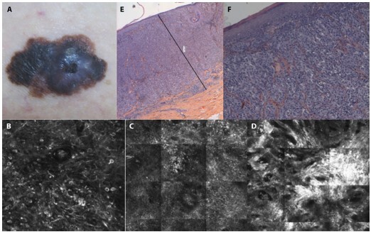
Results: Ninety-two primary melanomas were included: 44 males and 48 females (mean age 60.4 years, standard deviation [SD] 16.2) with a mean Breslow of 1.43 mm (SD 1.6). The most frequent dermoscopic presentation was the multicomponent pattern, the predominant confocal pattern was dendritic-cell type (44.6%). The presence of pigmented network on dermoscopy was related to lower Breslow and mitotic rates (both P = 0.002); in contrast to the presence of visible vessels, which was related to higher Breslow and mitotic indexes (both P = 0.001). Confocal observation of dermal nests or atypical cells in the papillary dermis was related to a higher mitotic rate (P = 0.006 and P = 0.03, respectively). Similarly, diffuse inflammatory infiltrates visible in the superficial dermis was associated with higher Breslow (P = 0.04) and mitotic index (P = 0.04).
Conclusions: Dermoscopic and RCM in vivo findings on primary melanoma correlate with histopathologic Breslow index, mitotic rate and tumor infiltrating lymphocytes. The architecture and cytology of primary melanoma can be estimated by combining dermoscopy and RCM prior to excision.
Keywords: Breslow index; confocal microscopy; melanoma; mitotix rate; prognostic markers.
©2022 Barragán-Estudillo et al.
Conflict of interest statement
Competing interests: None.
Figures
Similar articles
-
Role of In Vivo Reflectance Confocal Microscopy in the Analysis of Melanocytic Lesions.Acta Dermatovenerol Croat. 2018 Apr;26(1):64-67. Acta Dermatovenerol Croat. 2018. PMID: 29782304 Review.
-
Association Between Confocal Morphologic Classification and Clinical Phenotypes of Multiple Primary and Familial Melanomas.JAMA Dermatol. 2016 Oct 1;152(10):1099-1105. doi: 10.1001/jamadermatol.2016.1189. JAMA Dermatol. 2016. PMID: 27579522
-
Nevus-associated melanoma: An observational retrospective study of 22 patients evaluated with dermoscopy and reflectance confocal microscopy.Skin Res Technol. 2020 Jan;26(1):99-104. doi: 10.1111/srt.12770. Epub 2019 Sep 25. Skin Res Technol. 2020. PMID: 31556144
-
Histopathologic and Immunohistochemical Correlates of Confocal Descriptors in Pigmented Facial Macules on Photodamaged Skin.JAMA Dermatol. 2017 Aug 1;153(8):771-780. doi: 10.1001/jamadermatol.2017.1323. JAMA Dermatol. 2017. PMID: 28564685 Free PMC article.
-
Review of Dermoscopy and Reflectance Confocal Microscopy Features of the Mucosal Melanoma.Diagnostics (Basel). 2021 Jan 8;11(1):91. doi: 10.3390/diagnostics11010091. Diagnostics (Basel). 2021. PMID: 33429900 Free PMC article. Review.
Cited by
-
Optically Guided High-Frequency Ultrasound Shows Superior Efficacy for Preoperative Estimation of Breslow Thickness in Comparison with Multispectral Imaging: A Single-Center Prospective Validation Study.Cancers (Basel). 2023 Dec 28;16(1):157. doi: 10.3390/cancers16010157. Cancers (Basel). 2023. PMID: 38201584 Free PMC article.
-
Multiple Primary Melanoma: A Five-Year Prospective Single-Center Follow-Up Study of Two MC1R R/R Genotype Carriers.Life (Basel). 2023 Oct 23;13(10):2102. doi: 10.3390/life13102102. Life (Basel). 2023. PMID: 37895483 Free PMC article.
-
Spaghetti Technique Versus Wide Local Excision for Lentigo Maligna Affecting the Head and Neck Regions: Surgical Outcome and Descriptive Analysis of 79 Cases from a Single Practice Cohort.Dermatol Pract Concept. 2023 Jul 1;13(3):e2023193. doi: 10.5826/dpc.1303a193. Dermatol Pract Concept. 2023. PMID: 37557139 Free PMC article.
References
-
- Gershenwald JE, Scolyer RA, Hess KR, et al. Melanoma of the Skin. AJCC Cancer Staging Man. 2017:1–5. doi: 10.1001/jama.2010.1525. - DOI
LinkOut - more resources
Full Text Sources