Review
. 2023 May 22;119(5):1117-1129.
doi: 10.1093/cvr/cvac171.

Pathophysiology and pharmacology of G protein-coupled receptors in the heart

Affiliations
Review

Pathophysiology and pharmacology of G protein-coupled receptors in the heart

Alyssa Grogan et al. Cardiovasc Res. .

Abstract

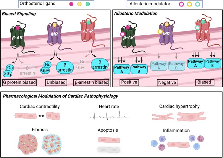
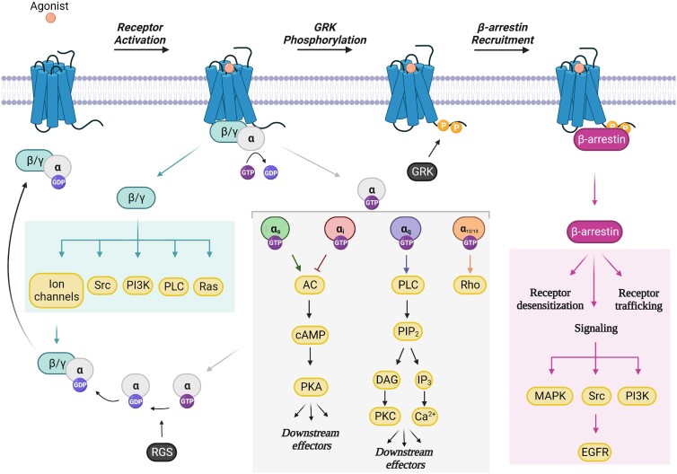
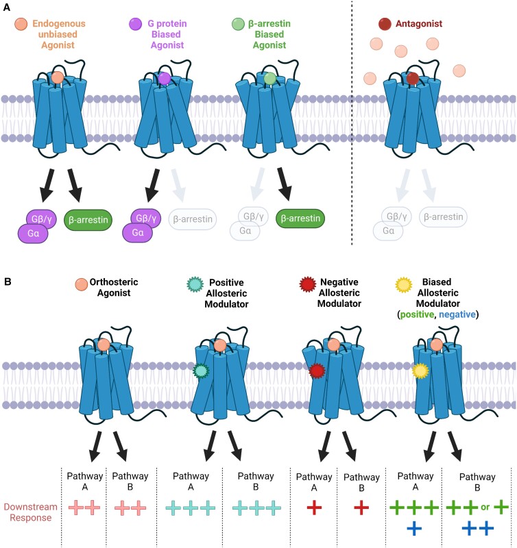
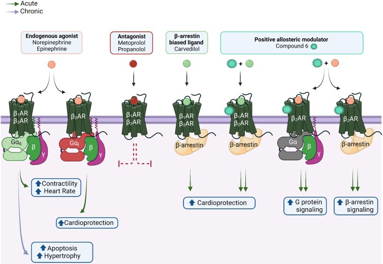
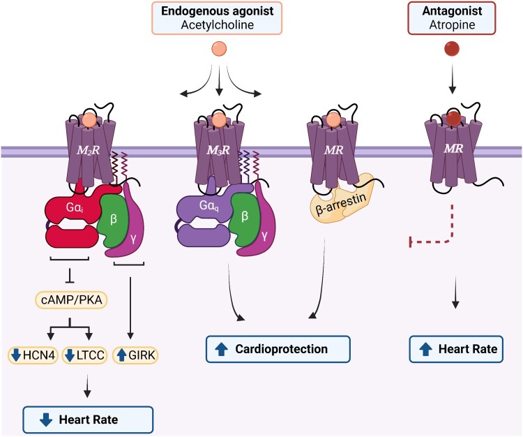
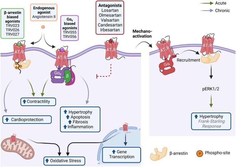
G protein-coupled receptors (GPCRs), comprising the largest superfamily of cell surface receptors, serve as fundamental modulators of cardiac health and disease owing to their key roles in the regulation of heart rate, contractile dynamics, and cardiac function. Accordingly, GPCRs are heavily pursued as drug targets for a wide variety of cardiovascular diseases ranging from heart failure, cardiomyopathy, and arrhythmia to hypertension and coronary artery disease. Recent advancements in understanding the signalling mechanisms, regulation, and pharmacological properties of GPCRs have provided valuable insights that will guide the development of novel therapeutics. Herein, we review the cellular signalling mechanisms, pathophysiological roles, and pharmacological developments of the major GPCRs in the heart, highlighting the β-adrenergic, muscarinic, and angiotensin receptors as exemplar subfamilies.

Keywords: Allosteric modulators; Biased signalling; G protein-coupled receptors; Heart failure.


Conflict of interest statement

Conflict of interest: H.A.R. is a scientific cofounder of Trevena Inc., a company that is developing new GPCR ligands.

Figures

Graphical Abstract
Graphical Abstract
Figure 1
Figure 1
GPCR activation and downstream signalling. Following agonist binding, GPCRs recruit heterotrimeric G proteins (Gα, β, γ) and induce the exchange of GDP for GTP on Gα. The Gα and Gβγ subunits subsequently dissociate forming two activated G protein units that independently transduce signals to downstream effectors. The four families of Gα (Gαs, Gαi, Gαq, and Gα12/13) activate distinct signalling cascades, thereby generating unique cellular responses. RGS proteins accelerate the GTPase activity of Gα, thereby leading to the inactivation of G protein signalling. GRK-mediated phosphorylation of the COOH-terminus of the receptor facilitates the recruitment of β-arrestin, which in turn, promotes receptor desensitization, receptor trafficking/recycling, and β-arrestin-dependent signalling mechanisms. GDP, guanosine diphosphate; GTP, guanosine triphosphate; PI3K, phosphoinositide 3-kinase; PLC, phospholipase C; AC, adenylyl cyclase; cAMP, cyclic adenosine monophosphate; PKA, protein kinase A; PIP2, phosphatidylinositol 4,5-bisphosphate; DAG, diacylglycerol; PKC, protein kinase C; IP3, inositol trisphosphate; MAPK, mitogen-activated protein kinase; EGFR, epidermal growth factor receptor; GRK, GPCR kinase; RGS, regulators of G protein signalling. Figure generated with Biorender.com.
Figure 2
Figure 2
Biased agonism and allosteric modulation of GPCR function. (A) Unbiased orthosteric agonists non-selectively activate G protein and β-arrestin signalling pathways, while biased orthosteric ligands preferentially activate one pathway over the other. Conversely, antagonists competitively inhibit agonist binding and thereby inactivate both G protein and β-arrestin signalling in a balanced fashion. (B) Allosteric modulators bind to sites that are topographically distinct from the orthosteric binding pocket of the receptor. Functionally, positive allosteric modulators enhance (+++) the activity of an orthosteric agonist, while negative allosteric modulators reduce (+) orthosteric activity. Biased allosteric modulators can preferentially enhance (positive; +++) or reduce (negative; +) the activity of a particular pathway (i.e. pathway A) while having no effect (++) and/or antagonizing (+) an alternative pathway (i.e. pathway B) in the presence of an orthosteric agonist. Figure generated with Biorender.com.
Figure 3
Figure 3
Physiological and pathological effects of cardiac βAR signalling. Norepinephrine and epinephrine, the endogenous βAR agonists, activate both β1AR and β2AR subtypes, albeit with different potencies. In general, β1AR is coupled to Gαs and β2AR is coupled to both Gαs and Gαi. In the short term, the activation of β1AR/β2AR through Gαs generates positive chronotropic and inotropic responses; however, chronic activation through Gαs can induce cardiomyocyte apoptosis and hypertrophy. In contrast, Gαi-mediated signalling via β2AR is thought to be cardioprotective due to its anti-apoptotic and anti-fibrotic effects. Furthermore, both receptor subtypes can couple to β-arrestin, which also induces cardioprotective signalling cascades in the heart. Carvedilol, a clinically utilized β-arrestin-biased ligand, preferentially stimulates β-arrestin signalling while antagonizing G protein signalling. Notably, carvedilol-mediated β-arrestin-dependent cardioprotection is potentiated by Compound 6, a positive allosteric modulator of the β1AR and β2AR. Additionally, in response to agonist, Compound 6 potentiates G protein and β-arrestin signalling mediated by β2AR, but not β1AR. As expected, traditional antagonists such as metoprolol or propranolol, block βAR signalling via both G protein and β-arrestin. Figure generated with biorender.com.
Figure 4
Figure 4
Physiological effects of cardiac MR signalling. The M2R and M3R, depicted herein as representative MR subtypes given that their physiological roles in the heart are the most extensively characterized, signal via distinct G protein classes. The M2R, coupled to Gαi, is the most abundant cardiac subtype and negatively regulates heart rate in response to the endogenous agonist, acetylcholine. In contrast, activation of M3R, coupled with Gαq, is considered cardioprotective due to its anti-arrhythmic/anti-fibrotic properties and protection against ischaemic injury. All MR subtypes couple to β-arrestin following agonist binding which initiates cardioprotective signalling cascades in the heart. In response to an antagonist, such as atropine, MR signalling is blocked and heart rate is increased. HCN4, hyperpolarization-activated cyclic nucleotide-gated 4 channel; LTCC, L-type Ca2+ channel; PKA, protein kinase A; GIRK, G protein-coupled inward rectifying K+ channel. Figure generated with biorender.com.
Figure 5
Figure 5
Physiological and pathological effects of cardiac AT1R signalling. The AT1R is a pleiotropic receptor that localizes to the cellular, mitochondrial, and nuclear membranes of cells. Ligands that preferentially stabilize the AT1R/β-arrestin complex (TRV023, TRV026, TRV027) increase the contractility of cardiomyocytes and have cardioprotective effects under long-term use. Likewise, ligands that preferentially stabilize the AT1R/Gαq complex (TRV055 and TRV056) increase the contractility of cardiomyocytes. However, chronic activation of this signalling pathway promotes cardiac hypertrophy, apoptosis, fibrosis, and inflammation. The endogenous ligand for the AT1R, Ang II, is unbiased towards both signalling pathways. AT1R antagonists such as Losartan, Olmesartan, and Valsartan, among others, stabilize the receptor in an inactive conformation thereby blocking the activation of any signalling cascade. Another activation mechanism of the AT1R is via membrane stretch. In turn, β-arrestin is recruited in a process mediated by Gαi and activates ERK1/2 phosphorylation regulating the Frank–Starling mechanism and inducing hypertrophy. Finally, the activation of the AT1R at mitochondrial or nuclear membranes induces oxidative stress and activates gene transcription. Figure generated with Biorender.com.

References

    1. Pierce KL, Premont RT, Lefkowitz RJ. Seven-transmembrane receptors. Nat Rev Mol Cell Biol 2002;3:639–650. - PubMed
    1. Sriram K, Insel PA. G protein-coupled receptors as targets for approved drugs: how many targets and how many drugs? Mol Pharmacol 2018;93:251–258. - PMC - PubMed
    1. Bjarnadottir TK, Gloriam DE, Hellstrand SH, Kristiansson H, Fredriksson R, Schioth HB. Comprehensive repertoire and phylogenetic analysis of the G protein-coupled receptors in human and mouse. Genomics 2006;88:263–273. - PubMed
    1. Wang J, Gareri C, Rockman HA. G-Protein-Coupled receptors in heart disease. Circ Res 2018;123:716–735. - PMC - PubMed
    1. Jiang H, Galtes D, Wang J, Rockman HA. G protein-coupled receptor signaling: transducers and effectors. Am J Physiol Cell Physiol 2022;323:C731–C748. - PMC - PubMed

Publication types

Substances