. 2022 Dec 19;13(1):7812.
doi: 10.1038/s41467-022-35498-6.

Endothelial cells regulate astrocyte to neural progenitor cell trans-differentiation in a mouse model of stroke

Affiliations

Endothelial cells regulate astrocyte to neural progenitor cell trans-differentiation in a mouse model of stroke

Wenlu Li et al. Nat Commun. .

Abstract

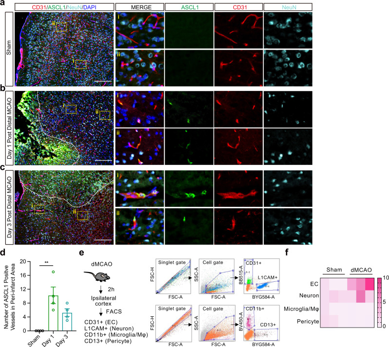
The concept of the neurovascular unit emphasizes the importance of cell-cell signaling between neural, glial, and vascular compartments. In neurogenesis, for example, brain endothelial cells play a key role by supplying trophic support to neural progenitors. Here, we describe a surprising phenomenon where brain endothelial cells may release trans-differentiation signals that convert astrocytes into neural progenitor cells in male mice after stroke. After oxygen-glucose deprivation, brain endothelial cells release microvesicles containing pro-neural factor Ascl1 that enter into astrocytes to induce their trans-differentiation into neural progenitors. In mouse models of focal cerebral ischemia, Ascl1 is upregulated in endothelium prior to astrocytic conversion into neural progenitor cells. Injecting brain endothelial-derived microvesicles amplifies the process of astrocyte trans-differentiation. Endothelial-specific overexpression of Ascl1 increases the local conversion of astrocytes into neural progenitors and improves behavioral recovery. Our findings describe an unexpected vascular-regulated mechanism of neuroplasticity that may open up therapeutic opportunities for improving outcomes after stroke.


Conflict of interest statement

The authors declare no competing interests.

Figures

Fig. 1
Fig. 1. Oxygen-glucose deprivation-stimulated brain endothelial cells convert astrocytes into neural progenitor cells.
a Morphologic changes of same astrocytes (AC) at day 0, day 1, day 2 and day 3 post cultured in conditioned neuron media collected from normal neurons (Neuron-con) or OGD-stimulated neurons (Neuron-ogd). b Morphologic changes of same AC at day 0, day 1, day 2 and day 3 post co-cultured with normal pericytes (PCcon) or OGD-stimulated pericytes (PCogd). c Morphologic changes of same AC at day 0, day 1, day 2 and day 3 post co-cultured with normal brain endothelial cells (ECcon) or OGD-stimulated brain endothelial cells (ECogd). d Representative immunostaining of DCX, PSA-NCAM and GFAP in AC or at day 3 post AC co-cultured with ECcon or ECogd. e mRNA levels of Nestin, DCX, TUJ1 and Glast at day 1 post AC co-cultured with ECcon or ECogd; n = 3 biologically independent samples; p < 0.0001. f mRNA levels of Nestin, DCX, TUJ1 and Glast at day 3 post AC co-cultured with ECcon or Ecogd; n = 3 biologically independent samples; p < 0.0001. g mRNA levels of Notch signaling-related genes at day 1 post AC co-cultured with ECcon or ECogd; n = 3 biologically independent samples. h mRNA levels of Notch signaling-related genes at day 3 post AC co-cultured with ECcon or ECogd; n = 3 biologically independent samples. p = 0.0077 for Notch1; p = 0.0017 for Dll1; p = 0.0007 for RBP-Jκ. Experiment was repeated independently 3 times with similar results (a–d). Data are shown as mean ± SEM. Two-way ANOVA with post-hoc Bonferroni adjustment. Scale bar, 50 μm. Source data are provided as a Source Data file.
Fig. 2
Fig. 2. Microvesicles derived from OGD-stimulated brain endothelial cells reprogram astrocytes into neural progenitor cells.
a Nanoparticle tracking analysis of microvesicles (MVs) derived from normal brain endothelial cells or OGD-stimulated brain endothelial cells; n = 3 biologically independent samples; p = 0.0354. b mRNA levels of Rab27a in normal brain endothelial cells or OGD-stimulated brain endothelial cells; n = 4 biologically independent samples; p = 0.0321. c Confocal microscopy revealed distribution of Cy3 labeled brain endothelial cells-derived MVs (EC-MVs) in AC at different time points. d Immunostaining images showing a red-to-green color switch of AC that had taken up Cre+ MVs derived from brain endothelial cells. e Representative immunostaining of DCX and GFAP in AC treated with vehicle or MVs derived from normal brain endothelial cells (ECcon-MVs) or OGD-stimulated brain endothelial cells (ECogd-MVs). f Quantification of DCX+ cells; n = 6 samples obtained from three independent experiments for vehicle group, n = 8 samples obtained from three independent experiments for ECcon-MVs group and ECogd-MVs group; Vehicle vs ECogd-MVs: p < 0.0001; ECcon-MVs vs ECogd-MVs: p = 0.0012. g Morphologic changes of same AC treated with ECogd-MVs at different time points. h Trans-differentiated cells in (g) were fixed at day 7 post ECogd-MVs treatment; Tuj1, Map2 and GFAP were detected by immunostaining. i Representative images of time-lapse live imaging analyses of individual AC after ECogd-MVs treatment. j Converted cells transduced with hSyn.GCaMP6S AAV and treated with 1 uM tetrodotoxin (TTX). k GCaMP6s traces for regions of interest 1–5; Arrows indicate the time points in (j). l Number of GCaMP+ cells in AC treated with vehicle or ECogd-MVs; n = 3 biologically independent samples for vehicle group, n = 4 biologically independent samples for ECogd-MVs group; p = 0.0217. Experiment was repeated independently 3 times with similar results (c, d, gj). Data are shown as mean ± SEM. Unpaired two-tailed t-test (a, b, l); one-way ANOVA with post-hoc Tukey adjustment (f). Scale bar, 25 μm (c, d); 50 μm (gj). Source data are provided as a Source Data file.
Fig. 3
Fig. 3. Brain endothelial cells-derived microvesicles containing pro-neural transcription factor Ascl1 initiate astrocytes trans-differentiation.
a Representative immunostaining of Ascl1 and GFAP at day 0, day 1 and day 2 post AC co-cultured with OGD-stimulated brain endothelial cells. b Protein levels of Ascl1 in AC or AC at day 2 post co-cultured with OGD-stimulated brain endothelial cells (ECogd); n = 4 biologically independent samples; p = 0.0173. c mRNA levels of Ascl1 in AC at day 0, day 1 and day 2 post co-cultured with OGD-stimulated brain endothelial cells; n = 3 biologically independent samples; p < 0.0001. d mRNA levels of Ascl1 in normal or OGD-stimulated brain endothelial cells; n = 4 biologically independent samples; p = 0.0471. e Protein levels of Ascl1 in MVs derived from normal or OGD-stimulated brain endothelial cells; n = 4 biologically independent samples; p = 0.0065. f Confocal microscopy revealed co-localization of GFP and Alix in brain endothelial cells transduced with GFP-Ascl1 lentivirus. g Experimental schematic for testing effects of MVs derived from brain endothelial cells overexpressed Ascl1 (ECas1-MVs) on AC. h Luciferase activity showed increased activities of Atoh8 and Hes6 in AC treated with ECas1-MVs; n = 4 biologically independent samples for Atoh8 group, n = 3 biologically independent samples for Hes6 group; p = 0.0265 for Atoh8; p = 0.0009 for Hes6. i Morphologic changes of same AC at day 0, day 1, day 3 and day 5 post ECas1-MVs treatment. j Representative immunostaining of DCX and Nestin in AC at day 3 post ECas1-MVs treatment. k Representative immunostaining of Map2 and Tuj1 in AC at day 7 post ECas1-MVs treatment; BF: bright filed. Experiment was repeated independently 3 times (a, f, ik) or 4 times (b, e) with similar results. Data are shown as mean ± SEM. Unpaired two-tailed t-test (b, d, e); one-way ANOVA with post-hoc Tukey adjustment (c); two-way ANOVA with post-hoc Bonferroni adjustment (h). Scale bar, 50 μm. Source data are provided as a Source Data file.
Fig. 4
Fig. 4. Pro-neural transcription factor Ascl1 is expressed in brain microvessels after focal cerebral ischemia in mice.
Representative immunostaining of Ascl1, CD31, and NeuN at shame (a), day 1 (b), and day 3 (c) post permanent distal middle cerebral artery occlusion (MCAO); Dashed white line indicates lesion border. d Vessels were counted in 0.5 mm2 peri-infarct ROIs per section (totally 3 sections per mouse, 4 mice per group; Criteria is any loci with more than one Ascl1 positive cell); p = 0.0027. e Isolation of brain endothelial cell, neuron, pericyte, and microglia/Mφ from ipsilateral cortex by fluorescence-activated cell sorting (FACS) at 2 h post permanent distal MCAO. f mRNA levels of Ascl1 in different cell isolated from (e). The experiment was repeated independently 4 times with similar results (a–c). Data are shown as mean ± SEM. One-way ANOVA with post-hoc Tukey adjustment. Scale bar, 200 μm. Source data are provided as a Source Data file.
Fig. 5
Fig. 5. Microvesicles derived from OGD-stimulated brain endothelial cells convert astrocytes into neural progenitor cells in mouse focal cerebral ischemia.
Confocal microscopy revealed Cy3 labeled primary brain endothelial cells-derived microvesicles (EC-MVs) could be taken into astrocytes at 3 h (a, b) and 24 h (a, c) post lateral ventricle injection; Dashed white line indicates lateral ventricle border. d Experimental design. e Representative immunostaining of GFP, GFAP, and NeuN at day 14 post lateral ventricle injection of AAV.GFA104.PI.eGFP. f Representative immunostaining of GFP and DCX at day 10 post 45 mins transient MCAO. g Number of DCX+ cells in 1 mm2 peri-infarct ROIs per section (totally 3 sections per mouse, 4 mice per group); p = 0.0026. h Number of both DCX+ and GFP+ cells in 1 mm2 peri-infarct ROIs per section (totally 3 sections per mouse, 4 mice per group); p = 0.0043. Experiment was repeated independently 3 times (a) or 4 times (e, f) with similar results. Data are shown as mean ± SEM. Unpaired two-tailed t-test. Scale bar, 25 μm (a); 200 μm (e, f). Source data are provided as a Source Data file.
Fig. 6
Fig. 6. Overexpression of Ascl1 in brain endothelial cells increases neurogenesis and improves neurological recovery after focal cerebral ischemia.
a AAV-FLEx-ASCL1-GFP or AAV-FLEx-GFP was injected into lateral ventricle of the Tie2-cre mice. Created with BioRender.com. b Representative immunostaining of GFP, CD31 and NeuN in cortex at day 8 post AAV-FLEx-ASCL1-GFP injection. c Representative immunostaining of GFP, CD31 and GFAP in cortex at day 8 post AAV-FLEx-ASCL1-GFP injection. d Experimental design. e Representative immunostaining of Ascl1 and TUJ1 at day 14 post permanent distal MCAO. f Quantification of TUJ+ cells expressing Ascl1 in the peri-infarct area (totally 3 sections per mouse, 4 mice per group); p = 0.0392. g Quantification of TUJ1+ cells in the peri-infarct area (totally 3 sections per mouse, 5 mice per group); p = 0.0496. h Representative immunostaining of Ascl1 and NeuN at day 14 post distal permanent MCAO; Dashed white line indicates lesion border. i Representative immunostaining of Ascl1 and NeuN at day 14 post distal permanent MCAO after AAV-FLEx-ASCL1-GFP injection. jk Quantification of NeuN+ cells (j) and Ascl1+ cells (k) in the peri-infarct area (totally 3 sections per mouse, 4 mice per group); p = 0.0245 in (j); p = 0.0323 in (k). l Foot fault test (n = 8 mice for AAV-FLEx-GFP group, n = 9 mice for AAV-FLEx-ASCL1-GFP group); p = 0.0439. The experiment was repeated independently 5 times with similar results (b, c). Data are shown as mean ± SEM. Unpaired two-tailed t-test (f, g, j, k); two-way ANOVA with post-hoc Bonferroni adjustment (l). Scale bar, 25 μm (e, i); 100 μm (b, c, h). Source data are provided as a Source Data file.

References

    1. Sharp FR, Liu J, Bernabeu R. Neurogenesis following brain ischemia. Brain Res. Dev. Brain Res. 2002;134:23–30. doi: 10.1016/S0165-3806(01)00286-3. - DOI - PubMed
    1. Hermann DM, Chopp M. Promoting brain remodelling and plasticity for stroke recovery: therapeutic promise and potential pitfalls of clinical translation. Lancet Neurol. 2012;11:369–380. doi: 10.1016/S1474-4422(12)70039-X. - DOI - PMC - PubMed
    1. Carmichael ST. Emergent properties of neural repair: elemental biology to therapeutic concepts. Ann. Neurol. 2016;79:895–906. doi: 10.1002/ana.24653. - DOI - PMC - PubMed
    1. Liang H, et al. Region-specific and activity-dependent regulation of SVZ neurogenesis and recovery after stroke. Proc. Natl Acad. Sci. USA. 2019;116:13621–13630. doi: 10.1073/pnas.1811825116. - DOI - PMC - PubMed
    1. Sun C, et al. Conditional ablation of neuroprogenitor cells in adult mice impedes recovery of poststroke cognitive function and reduces synaptic connectivity in the perforant pathway. J. Neurosci. 2013;33:17314–17325. doi: 10.1523/JNEUROSCI.2129-13.2013. - DOI - PMC - PubMed

Publication types