. 2023 Jan;30(1):31-37.
doi: 10.1038/s41594-022-00886-5. Epub 2022 Dec 19.

A pioneer factor locally opens compacted chromatin to enable targeted ATP-dependent nucleosome remodeling

Affiliations

A pioneer factor locally opens compacted chromatin to enable targeted ATP-dependent nucleosome remodeling

Megan A Frederick et al. Nat Struct Mol Biol. 2023 Jan.

Abstract

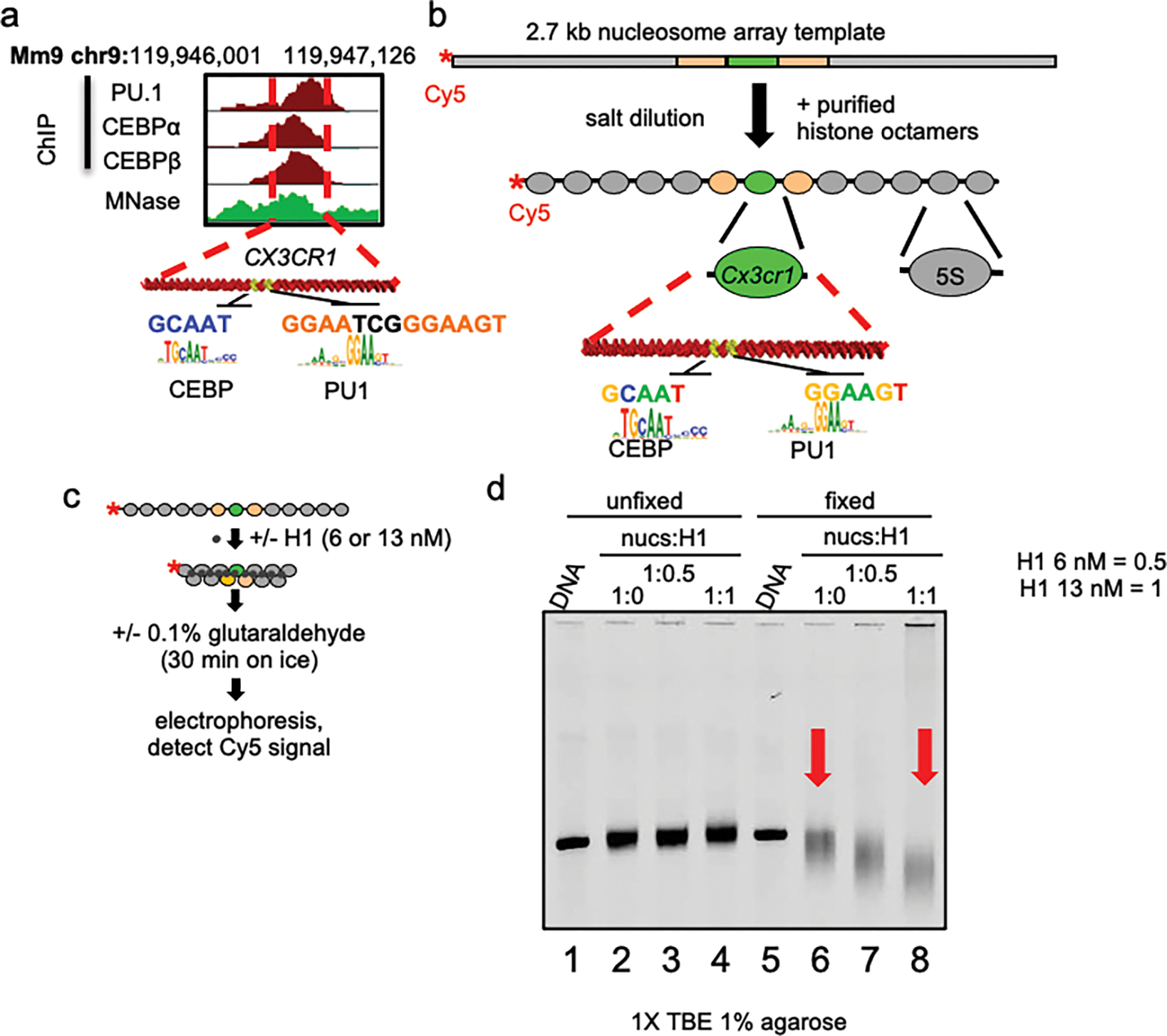
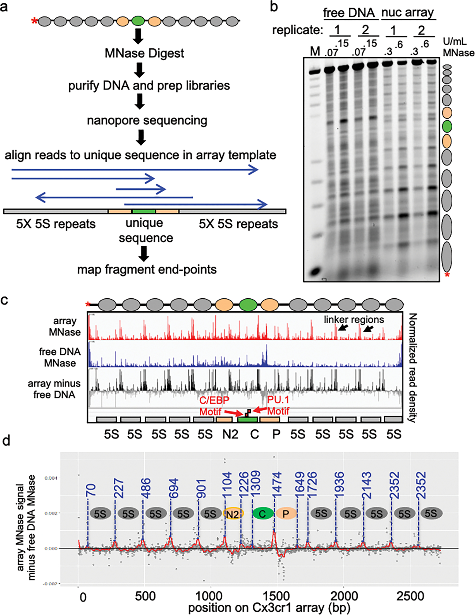
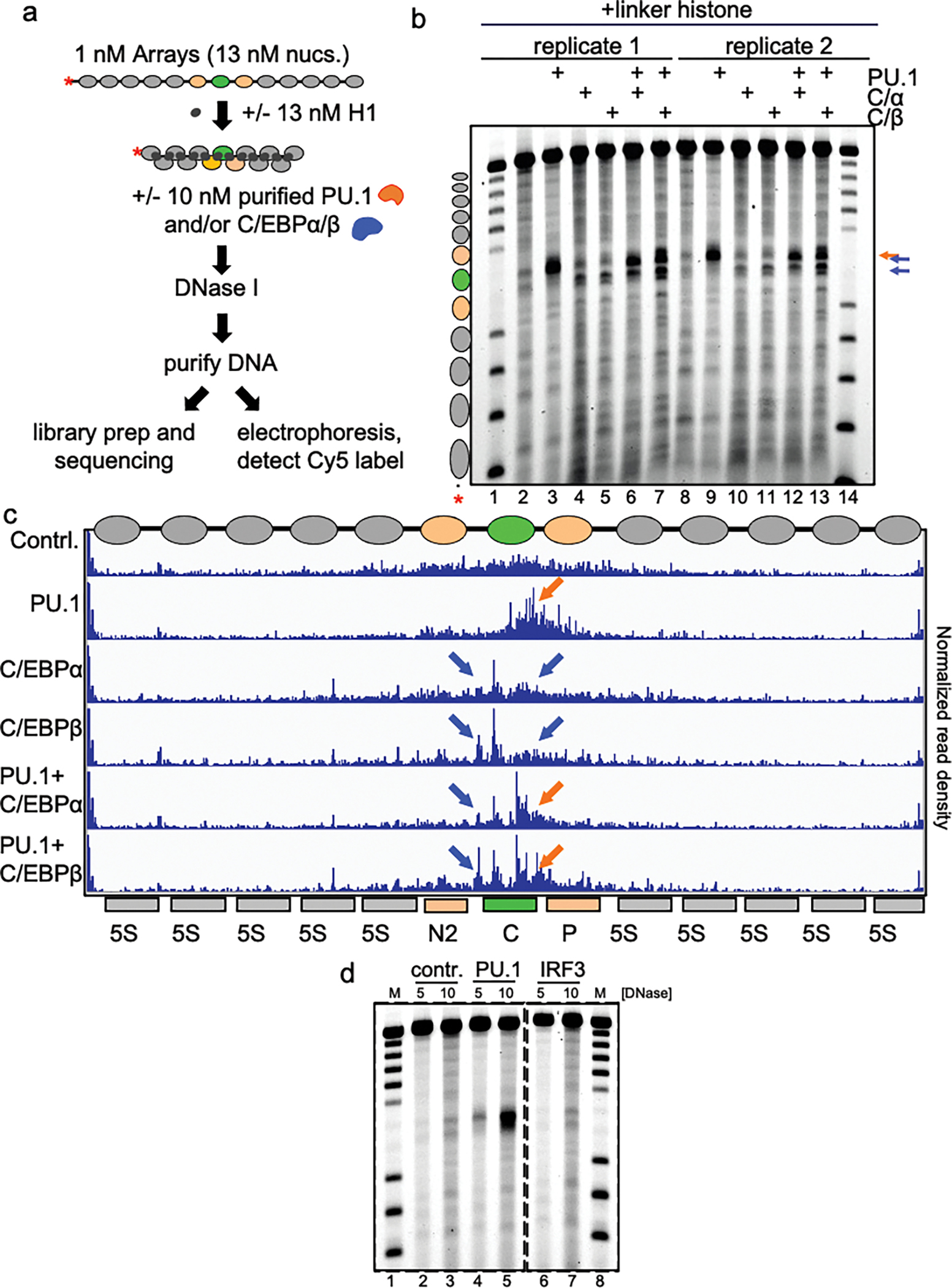
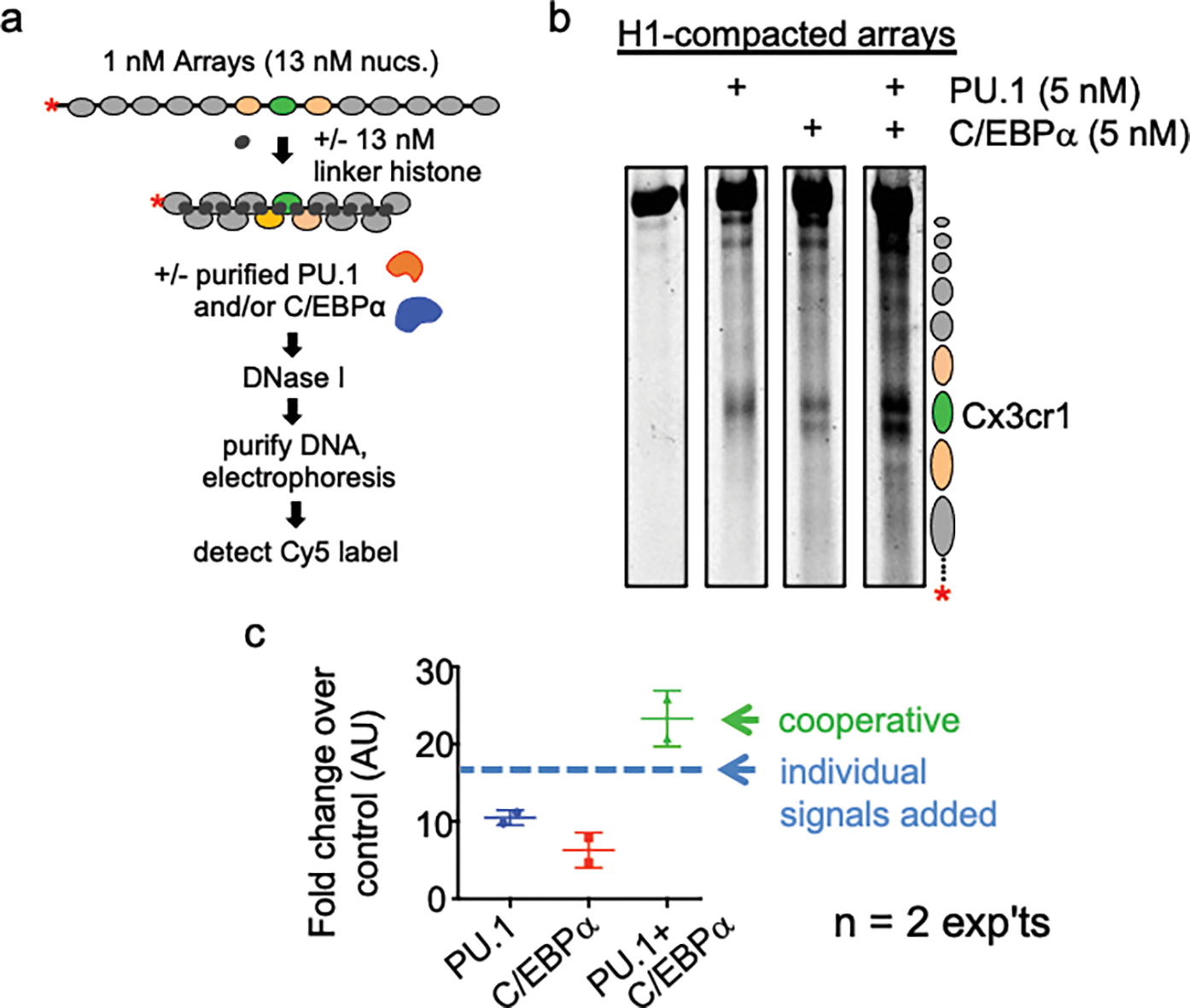
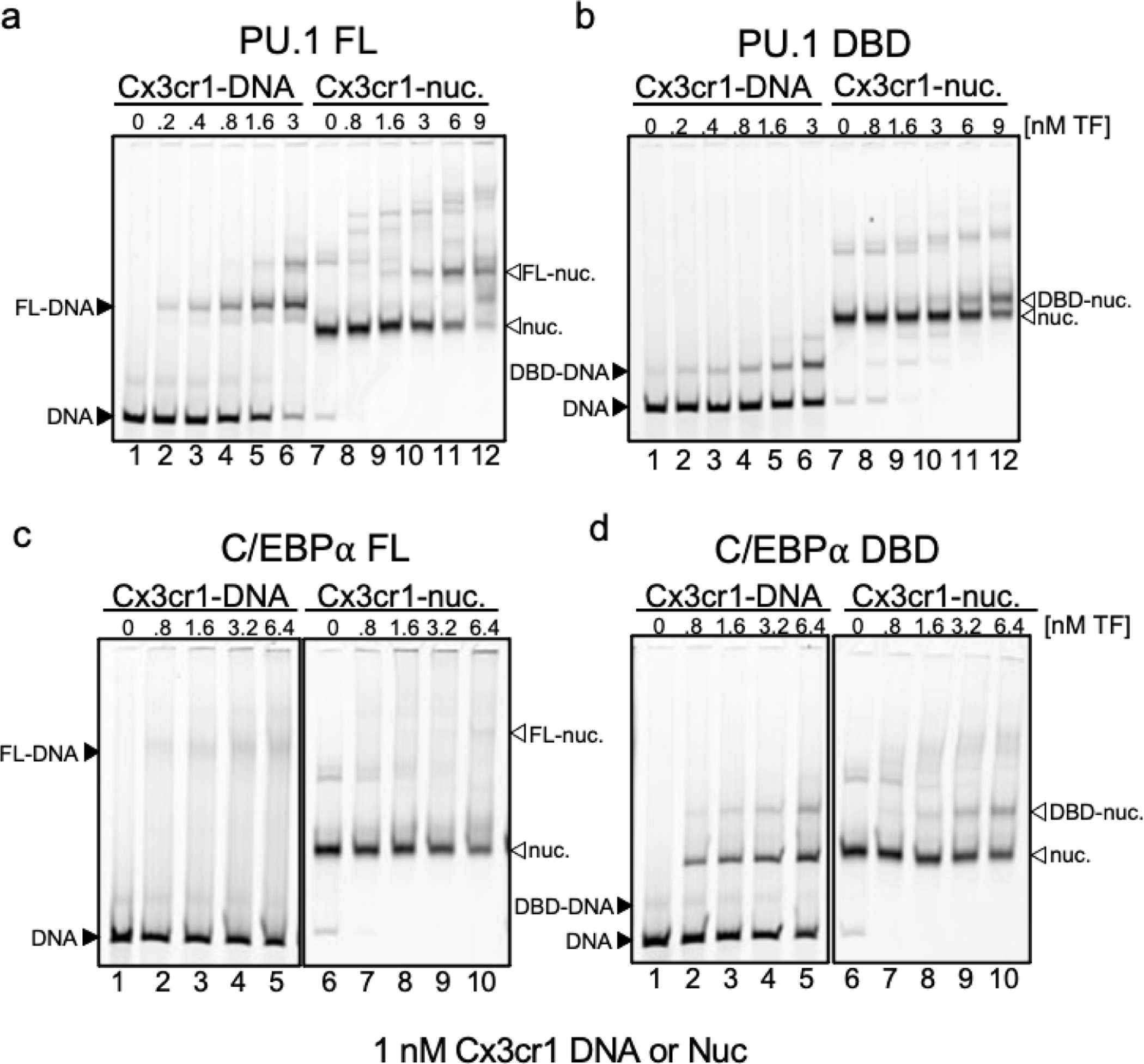
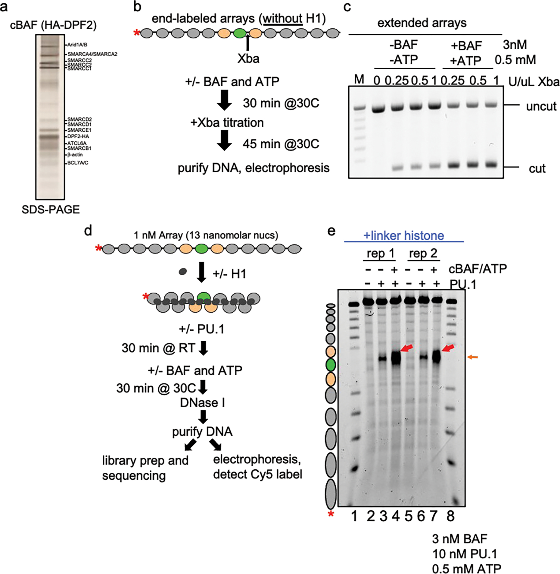
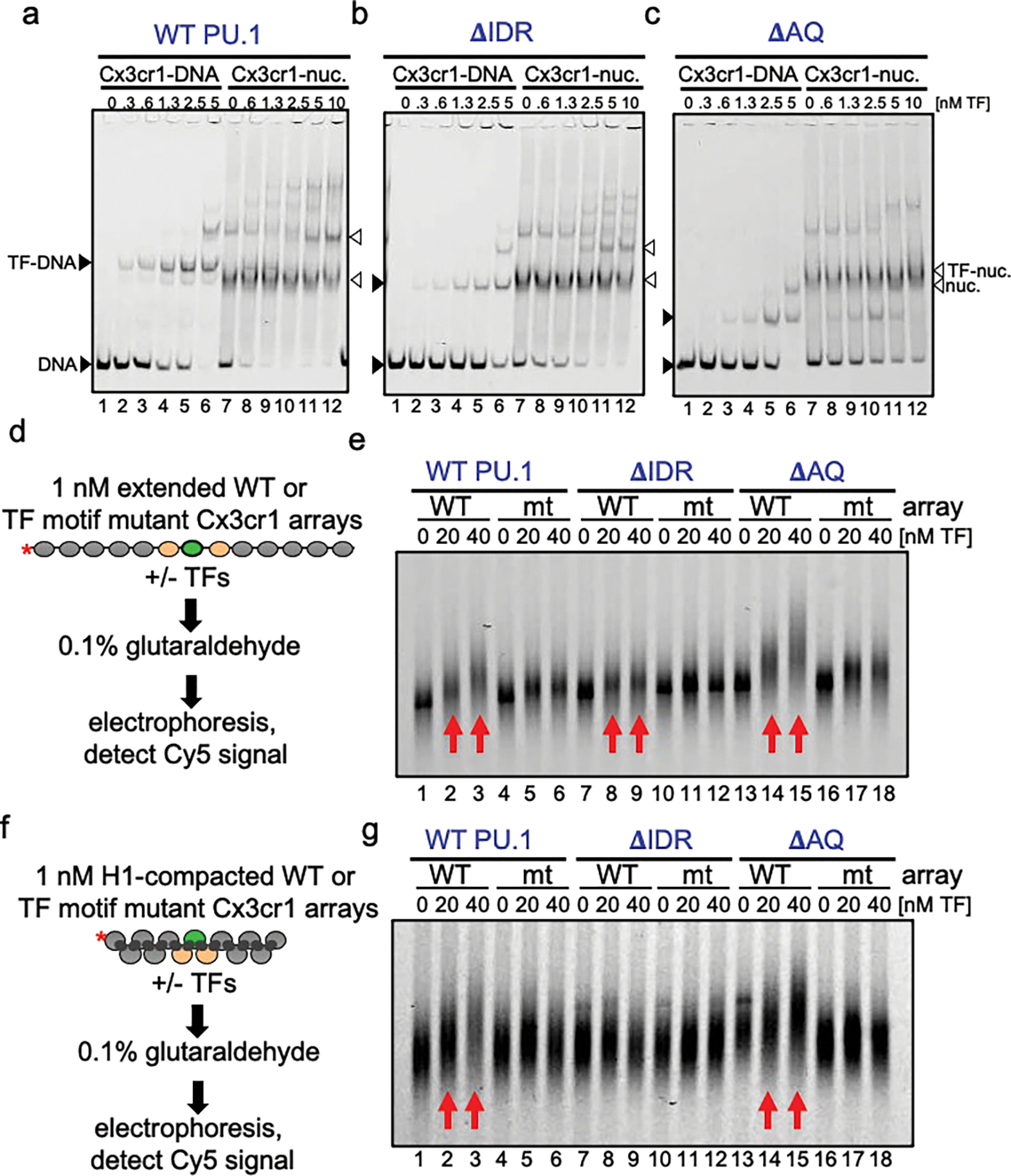
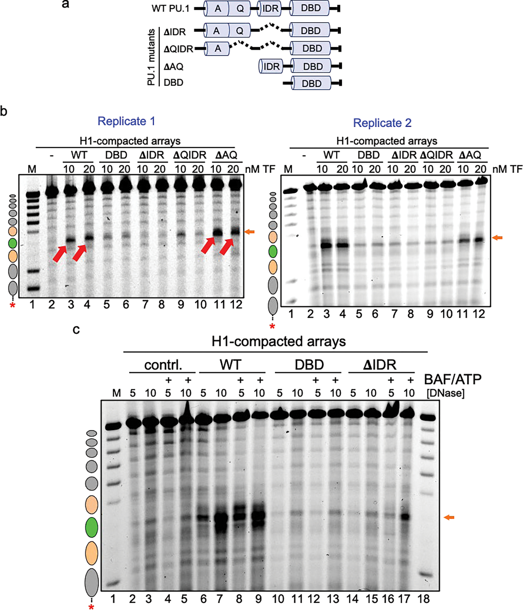
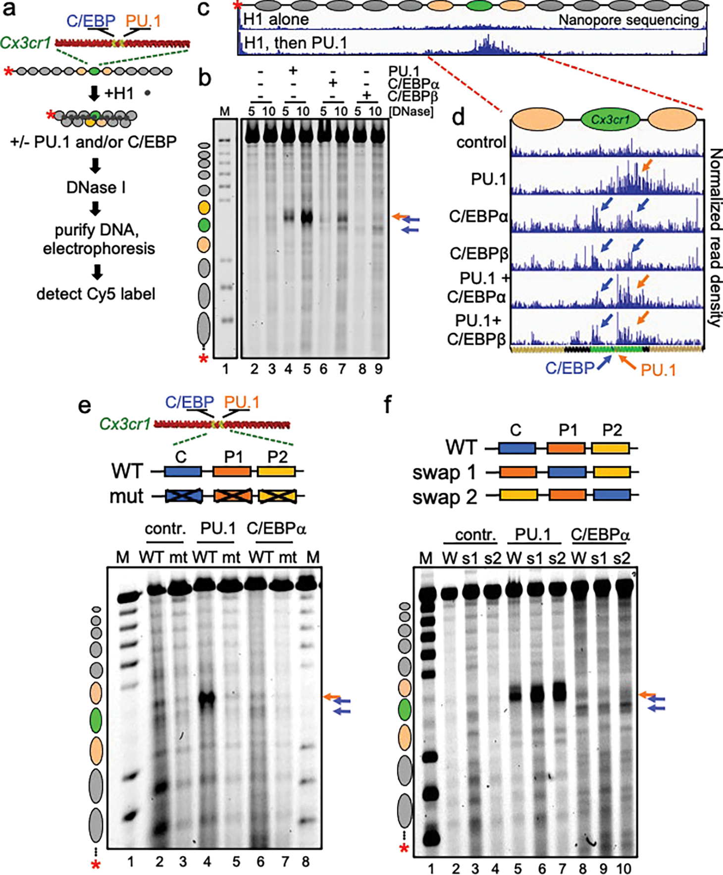
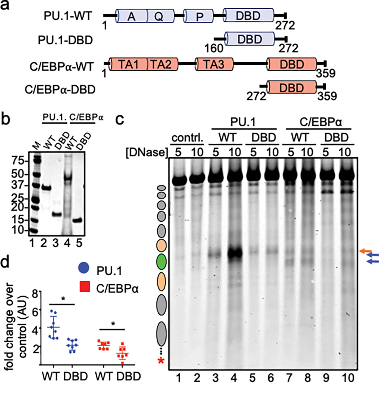
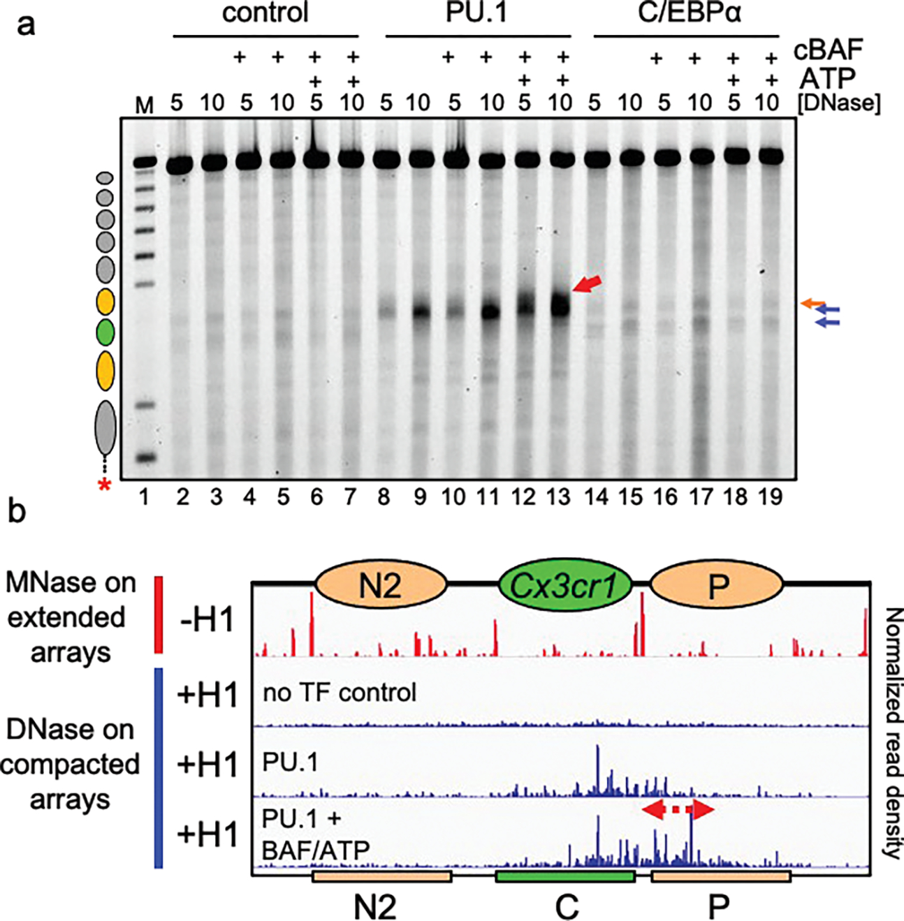
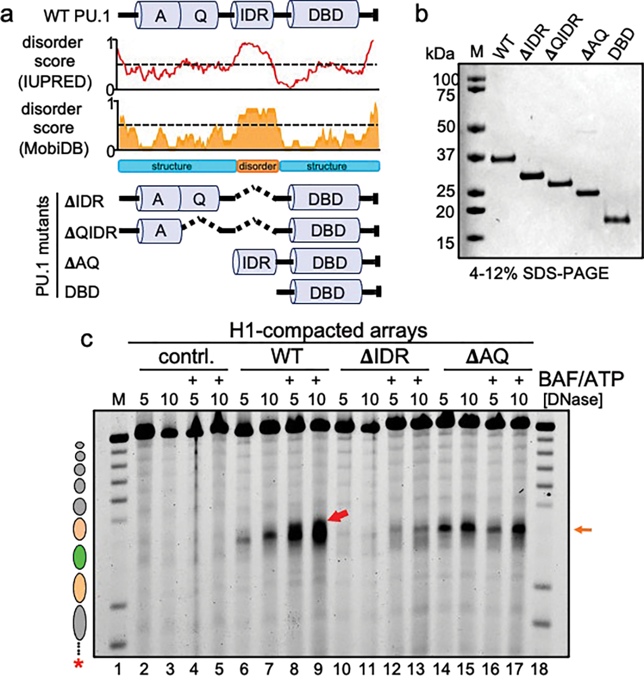
To determine how different pioneer transcription factors form a targeted, accessible nucleosome within compacted chromatin and collaborate with an ATP-dependent chromatin remodeler, we generated nucleosome arrays in vitro with a central nucleosome containing binding sites for the hematopoietic E-Twenty Six (ETS) factor PU.1 and Basic Leucine Zipper (bZIP) factors C/EBPα and C/EBPβ. Our long-read sequencing reveals that each factor can expose a targeted nucleosome on linker histone-compacted arrays, but with different nuclease sensitivity patterns. The DNA binding domain of PU.1 binds mononucleosomes, but requires an additional intrinsically disordered domain to bind and open compacted chromatin. The canonical mammalian SWI/SNF (cBAF) remodeler was unable to act upon two forms of locally open chromatin unless cBAF was enabled by a separate transactivation domain of PU.1. cBAF potentiates the PU.1 DNA binding domain to weakly open chromatin in the absence of the PU.1 disordered domain. Our findings reveal a hierarchy by which chromatin is opened and show that pioneer factors can provide specificity for action by nucleosome remodelers.


Conflict of interest statement

Competing interests: C.K. is the Scientific Founder, fiduciary Board of Directors member, Scientific Advisory Board member, shareholder, and consultant for Foghorn Therapeutics, Inc. C.K. also serves on the scientific advisory boards of Nereid Therapeutics, Nested Therapeutics, and is a consultant for Cell Signaling Technologies. K.E.W. is an employee and shareholder of Flare Therapeutics. The remaining authors declare no competing interest.

Figures

Extended Data Figure 1:
Extended Data Figure 1:. Generation of H1-compacted Cx3cr1 nucleosome arrays.
a, PU.1 and C/EBP ChIP-seq profiles (red) in macrophages and MNase-seq profile (green) in fibroblasts near the Cx3cr1 gene within the displayed region of the mouse genome with TF motifs indicated. b, Diagram of the dCTP-Cy5 end-labeled Cx3cr1 nucleosome array. c, Schematic of linker histone-mediated chromatin compaction. d, Analysis of linker histone-mediated chromatin compaction by native agarose gel electrophoresis. Free DNA (lanes 1 and 5) and nucleosome arrays with nucleosome:linker histone ratios of 0 (lanes 2 and 6), 0.5 (lanes 3 and 7) and 1.0 (lanes 4 and 8) are shown.
Extended Data Figure 2:
Extended Data Figure 2:. Nanopore sequencing to determine translational position of nucleosomes in the Cx3cr1 array.
a, Schematic of Nanopore sequencing and endpoint analysis pipeline. b, MNase digestion analysis of free DNA and extended nucleosome arrays at two MNase concentrations (U/mL) visualized by gel electrophoresis. Reactions shown in this gel are the same ones analyzed by nanopore sequencing. c, IGV visualization of Nanopore sequencing endpoint analysis of MNase digested free DNA (0.07 U/mL MNase), extended nucleosome arrays (0.3 U/mL) and free DNA signal subtracted from array signal to account for MNase site bias. Plots show normalized read density on the y axis. For each plot, the maximum value is set to 0.4% of reads. d, Determination of nucleosome translational positions.
Extended Data Figure 3:
Extended Data Figure 3:. Nanopore sequencing of TFs incubated with H1-compacted arrays.
a, Schematic of DNase I digestion analysis. b, DNase I digestion analysis of TFs binding to H1-compacted nucleosome arrays visualized by gel electrophoresis (10 ng/uL DNase I). The same samples shown on the gel were used for nanopore sequencing shown in panel c. c, IGV visualization of Nanopore endpoint analysis of DNase I digested H1-compacted nucleosome arrays incubated with indicated transcription factor(s). Plots show normalized read density on the y axis. For each plot, the maximum value is set to 0.4% of reads. d, DNase I digestion analysis of indicated TFs binding to H1-compacted nucleosome arrays visualized by gel electrophoresis.
Extended Data Figure 4:
Extended Data Figure 4:. PU.1 and C/EBPa open chromatin cooperatively.
a, Schematic of DNase I digestion analysis. b, DNase I digestion analysis of TFs binding to H1-compacted nucleosome arrays visualized by gel electrophoresis. c, Quantified Cy5 signal in each lane normalized to no TF control (n=2).
Extended Data Figure 5:
Extended Data Figure 5:. DNA-binding domains of PU.1 and C/EBPα are sufficient for nucleosome binding.
a-d, Representative EMSAs showing the affinity of increasing amounts of a, Full-length PU.1 (PU.1 FL), b, PU.1-DBD, c, Full-length C/EBPα (C/EBPα FL), d, C/EBP-DBD to Cy5-labeled Cx3cr1 free DNA (black arrows) or mononucleosomes (white arrows).
Extended Data Figure 6:
Extended Data Figure 6:. cBAF readily remodels extended nucleosome arrays but requires PU.1 to access H1-compacted arrays.
a, silver-stain of affinity-purified cBAF complexes from mammalian HEK293T cells expressing HA-DPF2. b, Schematic of XbaI accessibility assay with extended arrays and cBAF remodeling complex. c, Agarose gel visualization of XbaI accessibility assay of extended nucleosome arrays (no linker histone) incubated without and with cBAF plus ATP. d, Schematic of DNase I digestion analysis of transcription factors and cBAF with H1-compacted arrays. e, DNase I digestion analysis H1-compacted nucleosome arrays incubated alone (no TF, lanes 2 and 5), PU.1 alone (lanes 3 and 6), and PU.1 with cBAF and ATP (lanes 4 and 7). The same samples were used to perform nanopore sequencing shown in Fig. 3b.
Extended Data Figure 7:
Extended Data Figure 7:. Hierarchy of chromatin binding by PU.1 WT and mutants.
a-c, Representative EMSAs showing the affinity of increasing amounts of a, WT PU.1, b, ΔIDR, c, ΔAQ to Cy5-labeled Cx3cr1 free DNA (black arrows) or mononucleosomes (white arrows). d, Schematic of EMSA performed with transcription factors and extended nucleosome arrays e, EMSA showing the affinity of increasing amounts of WT PU.1 (lanes 1–6), ΔIDR (lanes 7–12), and ΔAQ (lanes 13–18) to Cy5-labeled extended WT or TF motif mutant Cx3cr1 nucleosome arrays. f, Schematic of EMSA performed with transcription factors and H1-compacted nucleosome arrays. g, Representative EMSA showing the affinity of increasing amounts of WT PU.1 (lanes 1–6), ΔIDR (lanes 7–12), and ΔAQ (lanes 13–18) to Cy5-labeled H1-compacted WT or TF motif mutant Cx3cr1 nucleosome arrays.
Extended Data Figure 8:
Extended Data Figure 8:. The IDR domain of PU.1 is most crucial for chromatin opening.
a, Illustration of the PU.1 deletion mutant series. Shown are the positions of the acidic domain (A), Q-rich domain (Q), intrinsically disordered region (IDR) and DNA-binding domain (DBD). b, DNase I digestion analysis of two transcription factor concentrations binding to H1-compacted nucleosome arrays visualized by gel electrophoresis (10 ng/uL DNase I). c, DNase I digestion analysis of H1-compacted nucleosome arrays incubated with no TFs (contrl., lanes 2–5), WT PU.1 (lanes 6–9), DBD (lanes 10–13), and ΔIDR deletion (lanes 14–17) with or without the addition of cBAF and ATP.
Figure 1:
Figure 1:. Different patterns of chromatin opening by PU.1 and C/EBP.
a, Schematic of chromatin assembly, compaction, transcription factor binding, and DNase I digestion assays. b, DNase I digestion analysis of TFs binding to H1-compacted nucleosome arrays visualized by gel electrophoresis at two DNase I concentrations (in ng/uL) Distinct hypersensitive sites are observed, relative to no TF control (lanes 2–3), upon PU.1 (lanes 4–5, orange arrow), C/EBPα (lanes 6–7, blue arrows), and C/EBPβ (lanes 8–9, blue arrows). Lane M, partial EcoRI digest of end-labeled array fragment. Positions of nucleosomes on reconstituted arrays are indicated. c and d, Nanopore sequencing endpoint analysis of DNase I digested (10 ng/uL) H1-compacted arrays. Ovals indicate the translational positions of nucleosomes determined in Extended Data Fig. 2d. Plots show normalized read density on the y axis. For each plot, the maximum value is set to 0.4% of reads. e, DNase I digestion (10 ng/uL) of H1-compacted wild-type Cx3cr1 nucleosome arrays (WT) or motif mutant Cx3cr1 nucleosome arrays (mt) incubated alone (contr., lanes 2–3), with PU.1 (lanes 4–5) or C/EBPα (lanes 6–7). f, DNase I digestion of H1-compacted wild-type Cx3cr1 nucleosome arrays (WT) or PU.1 and C/EBPα motif swap arrays (swap 1 or swap 2) incubated alone (contr., lanes 2–4), with PU.1 (lanes 5–7) or C/EBPα (lanes 8–10).
Figure 2:
Figure 2:. Domains outside of DBDs are needed to open chromatin.
a, Illustration of wild-type (WT) and DNA-binding domain (DBD) recombinant PU.1 and C/EBPα. b, Coomassie stained SDS-PAGE of 1 ug of each indicated purified protein. c, DNase I digestion analysis of WT and DBD TF opening of H1-compacted chromatin. d, Quantified Cy5 signal in each lane normalized to no TF control (n=4, *p < 0.05). Data are mean ± SD and significance was determined by Student’s unpaired t-test.
Figure 3:
Figure 3:. PU.1 Enables further chromatin opening by cBAF.
a, DNase I digestion analysis H1-compacted nucleosome arrays incubated with PU.1 (lanes 8–13) or C/EBPα (lanes 14–19), in the presence of cBAF alone (lanes 10–11 and lanes 16–17, respectively) or cBAF and ATP (lanes 12–13 and lanes 18–19, respectively). b, Nanopore analysis of MNase digested extended arrays (−H1) and DNase I digested compacted arrays (+H1) alone (no TF control), with PU.1 alone, or with PU.1, cBAF and ATP. Ovals indicate the translational positions of nucleosomes determined in Extended Data Fig. 2d. Plots show normalized read density on the y axis. For each plot, the maximum value is set to 0.4% of reads.
Figure 4:
Figure 4:. cBAF action requires AQ and IDR domains of PU.1.
a, Predicted disorder tendency of WT PU.1. The red and orange lines indicate the predicted disorder tendency along the WT PU.1 protein (as calculated by IUPred and MobiDB, respectively) with values above 0.5 (indicated by the dotted line) considered disordered. Shown are the positions of the Acidic domain (A), Q-rich domain (Q), Intrinsically disordered region (IDR) and DNA-binding domain (DBD). The deletion mutant series of PU.1 are indicated below as ΔIDR, ΔQIDR, ΔAQ, and DBD. b, SDS-PAGE analysis of 1 μg of each protein, stained with Coomassie blue. c, DNase I digestion analysis H1-compacted nucleosome arrays incubated alone (contrl., lanes 2–5), WT PU.1 (lanes 6–9), ΔIDR deletion (lanes 10–13), and ΔAQ truncation (lanes 14–17) with or without the addition of BAF and ATP.

References

    1. Iwafuchi-Doi M & Zaret KS Pioneer transcription factors in cell reprogramming. Genes and Development (2014) doi:10.1101/gad.253443.114. - DOI - PMC - PubMed
    1. Schwarz PM & Hansen JC Formation and stability of higher order chromatin structures Contributions of the histone octamer. Journal of Biological Chemistry (1994). - PubMed
    1. Adams CC & Workman JL Binding of disparate transcriptional activators to nucleosomal DNA is inherently cooperative. Molecular and Cellular Biology (2015) doi:10.1128/mcb.15.3.1405. - DOI - PMC - PubMed
    1. Mirny LA Nucleosome-mediated cooperativity between transcription factors. Proceedings of the National Academy of Sciences (2010) doi:10.1073/pnas.0913805107. - DOI - PMC - PubMed
    1. Bustin M, Catez F & Lim JH The dynamics of histone H1 function in chromatin. Molecular Cell (2005) doi:10.1016/j.molcel.2005.02.019. - DOI - PubMed

Publication types