. 2022 Dec 19;13(1):525.
doi: 10.1186/s13287-022-03208-0.

3D printing of injury-preconditioned secretome/collagen/heparan sulfate scaffolds for neurological recovery after traumatic brain injury in rats

Affiliations

3D printing of injury-preconditioned secretome/collagen/heparan sulfate scaffolds for neurological recovery after traumatic brain injury in rats

Xiao-Yin Liu et al. Stem Cell Res Ther. .

Abstract

Background: The effects of traumatic brain injury (TBI) can include physical disability and even death. The development of effective therapies to promote neurological recovery is still a challenging problem. 3D-printed biomaterials are considered to have a promising future in TBI repair. The injury-preconditioned secretome derived from human umbilical cord blood mesenchymal stem cells showed better stability in neurological recovery after TBI. Therefore, it is reasonable to assume that a biological scaffold loaded with an injury-preconditioned secretome could facilitate neural network reconstruction after TBI.

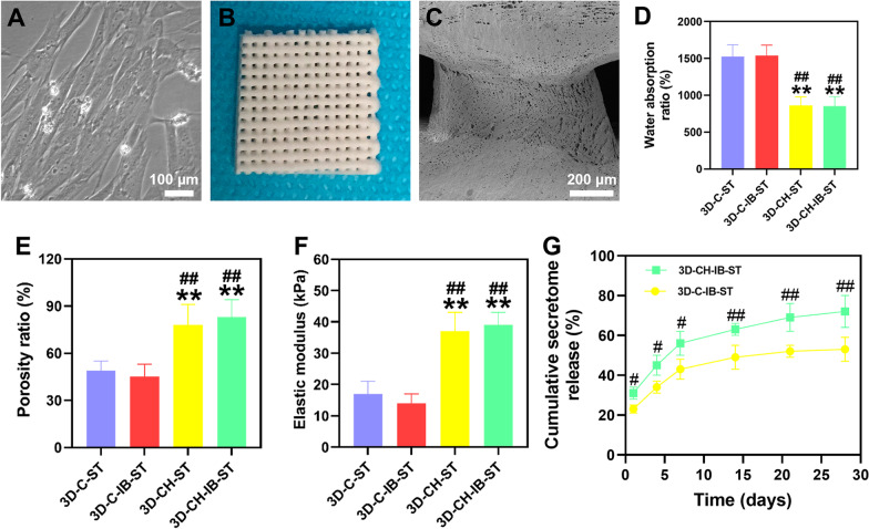
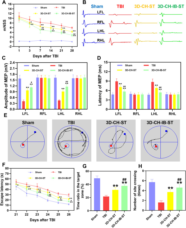
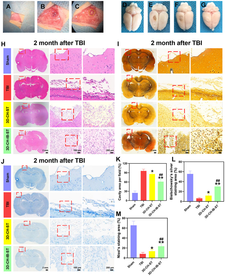
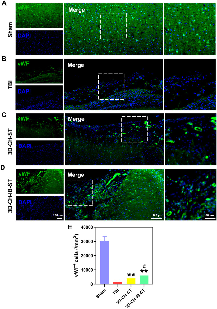
Methods: In this study, we fabricated injury-preconditioned secretome/collagen/heparan sulfate scaffolds by 3D printing. The scaffold structure and porosity were examined by scanning electron microscopy and HE staining. The cytocompatibility of the scaffolds was characterized by MTT analysis, HE staining and electron microscopy. The modified Neurological Severity Score (mNSS), Morris water maze (MWM), and motor evoked potential (MEP) were used to examine the recovery of cognitive and locomotor function after TBI in rats. HE staining, silver staining, Nissl staining, immunofluorescence, and transmission electron microscopy were used to detect the reconstruction of neural structures and pathophysiological processes. The biocompatibility of the scaffolds in vivo was characterized by tolerance exposure and liver/kidney function assays.

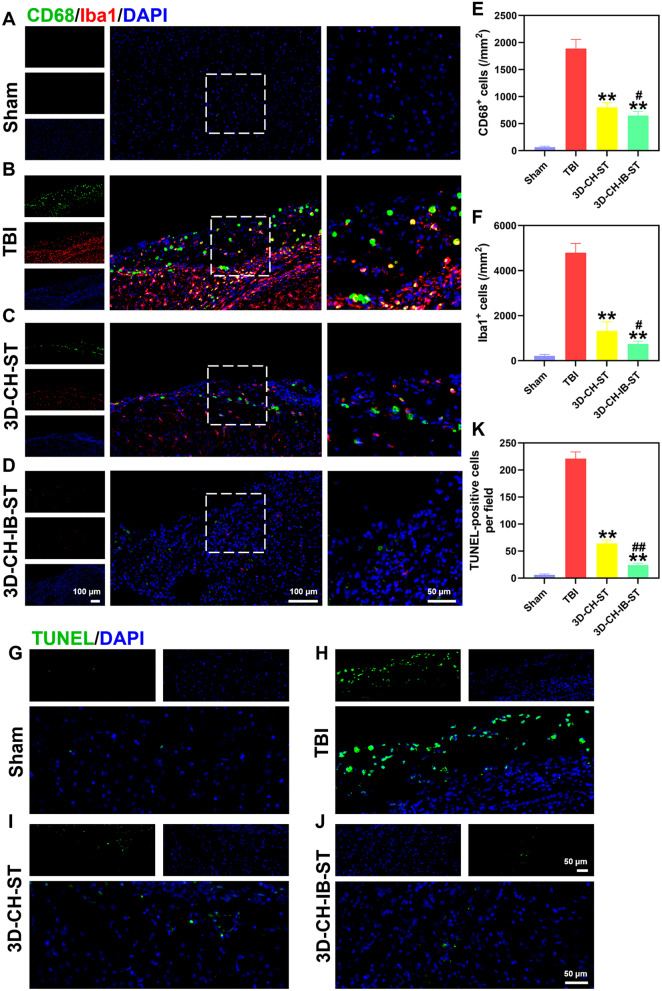
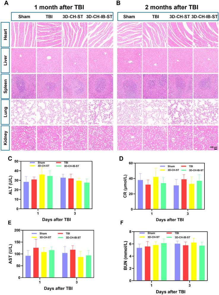
Results: The excellent mechanical and porosity characteristics of the composite scaffold allowed it to efficiently regulate the secretome release rate. MTT and cell adhesion assays demonstrated that the scaffold loaded with the injury-preconditioned secretome (3D-CH-IB-ST) had better cytocompatibility than that loaded with the normal secretome (3D-CH-ST). In the rat TBI model, cognitive and locomotor function including mNSS, MWM, and MEP clearly improved when the scaffold was transplanted into the damage site. There is a significant improvement in nerve tissue at the site of lesion. More abundant endogenous neurons with nerve fibers, synaptic structures, and myelin sheaths were observed in the 3D-CH-IB-ST group. Furthermore, the apoptotic response and neuroinflammation were significantly reduced and functional vessels were observed at the injury site. Good exposure tolerance in vivo demonstrated favorable biocompatibility of the scaffold.

Conclusions: Our results demonstrated that injury-preconditioned secretome/collagen/heparan sulfate scaffolds fabricated by 3D printing promoted neurological recovery after TBI by reconstructing neural networks, suggesting that the implantation of the scaffolds could be a novel way to alleviate brain damage following TBI.

Keywords: 3D printing; Biomaterial scaffolds; Human umbilical cord blood mesenchymal stem cells; Injury-preconditioned secretome; Neural reconstruction; Traumatic brain injury.


Conflict of interest statement

The authors declare that they have no competing interests.

Figures

Fig. 1
Fig. 1
A Morphology of HUCMSCs under phase contrast microscopy. B General view of 3D-CH-IB-ST. C Typical representative images of TEM (C) of 3D-CH-IB-ST. DF The water absorption (D), porosity ratio (E), and elastic modulus (F) of the 3D-C-ST and 3D-CH-IB-ST scaffolds. G Release profile of the secretome in 3D-C-IB-ST and 3D-CH-IB-ST for 30 days. **P < 0.01 versus the 3D-C-ST group, #P < 0.05, ##P < 0.01 versus the 3D-C-IB-ST group
Fig. 2
Fig. 2
AF Representative images of HUCMSCs cultured with 3D-CH-IB-ST scaffolds by phase contrast microscopy (A, B), SEM (C, D) and HE staining (E, F). G MTT analysis was performed after 1, 3, 5, and 7 days of HUCMSC cultured with 3D-CH-IB-ST. H, I MTT analysis H and cell adhesion rate I of NSCs cultured with 3D-CH-IB-ST. JQ Immunofluorescence staining of NF, GAP43, Tuj-1, and NeuN in NSCs cultured with 3D-CH-IB-ST. *P < 0.05, **P < 0.01 versus 3D-CH-ST group
Fig. 3
Fig. 3
A Rat mNSS scores were recorded at 1, 3, 7, 14, 21, and 28 days after implantation of differentiated scaffolds after TBI. B Representative motor evoked potential waveforms at 2 months posttransplant in the four groups. C, D The amplitude (C) and latency (D) of MEP in four groups. E Representative swimming paths in the Morris water maze in the four groups. FH Escape latency (F), time spent in the target quadrant (G), and number of site crossings (H) in the four groups. *P < 0.05, **P < 0.01 versus TBI group, #P < 0.05, ##P < 0.01 versus 3D-CH-ST group
Fig. 4
Fig. 4
AC Flowchart of 3D-CH-IB-ST implantation. DG Representative general views at 2 months after implantation in the four groups. HJ Representative images of HE staining (H), Bielschowsky’s silver staining (I), and Nissl staining (J) at 2 months after implantation in the four groups. KM Statistical calculation of the cavity area (K), Bielschowsky’s silver staining (L), and Nissl staining area (M) in the four groups. *P < 0.05, **P < 0.01 versus TBI group, ##P < 0.01 versus 3D-CH-ST group
Fig. 5
Fig. 5
AD The expression of Nestin around the injury site after TBI in the four groups. E Statistical analysis of Nestin+ cell numbers. FI The expression of NF, MBP, and NeuN around the injury site after TBI in the four groups. JL Statistical analysis of NF+ (J), MBP+ (K), and NeuN+ (L) cell numbers. **P < 0.01 versus TBI group, #P < 0.05, ##P < 0.01 versus 3D-CH-ST group
Fig. 6
Fig. 6
AD The expression of MAP2 and SYP around the injury site after TBI in the four groups. E, F Statistical analysis of MAP2+ (E) and SYP+ (F) cell numbers. GJ Representative TEM images of the four groups. KM Statistical analysis of the number of myelinated axons per 1000 μm2 (K), myelinated axon diameter (L), and myelin sheath thickness (M) in the four groups. *P < 0.05, **P < 0.01 versus TBI group, #P < 0.05, ##P < 0.01 versus 3D-CH-ST group
Fig. 7
Fig. 7
AD vWF expression around the injury site after TBI in the four groups. E Statistical analysis of vWF+ cell numbers. **P < 0.01 versus TBI group, #P < 0.05 versus 3D-CH-ST group
Fig. 8
Fig. 8
AD CD68/Iba1 expression around the injury site after TBI in the four groups. E, F Quantitative analysis of CD68+ (E) and Iba1+ (F) cell numbers. GJ Representative TUNEL staining in the four groups. Apoptotic cells labeled with TUNEL emitted green fluorescence. K Quantitative counting of TUNEL-positive cells. **P < 0.01 versus TBI group, #P < 0.05, ##P < 0.01 versus 3D-CH-ST group
Fig. 9
Fig. 9
A, B Representative HE staining images of major organs at 1 and 2 months after TBI. CF The levels of ALT, CR, AST, and BUN in plasma at 1 and 3 days after TBI

Similar articles

Cited by

References

    1. Kim JA, Sheth KN. Holding out hope after traumatic brain injury. JAMA Neurol. 2021;78(5):522–524. doi: 10.1001/jamaneurol.2020.5328. - DOI - PubMed
    1. Ashina H, Eigenbrodt AK, Seifert T, Sinclair AJ, Scher AI, Schytz HW, et al. Post-traumatic headache attributed to traumatic brain injury: classification, clinical characteristics, and treatment. Lancet Neurol. 2021;20(6):460–469. doi: 10.1016/S1474-4422(21)00094-6. - DOI - PubMed
    1. Afshari FT, Kappagantula S, Fawcett JW. Extrinsic and intrinsic factors controlling axonal regeneration after spinal cord injury. Expert Rev Mol Med. 2009;11:e37. doi: 10.1017/S1462399409001288. - DOI - PubMed
    1. Igarashi M, Takeuchi K, Sugiyama S. Roles of CSGalNAcT1, a key enzyme in regulation of CS synthesis, in neuronal regeneration and plasticity. Neurochem Int. 2018;119:77–83. doi: 10.1016/j.neuint.2017.10.001. - DOI - PubMed
    1. Shi W, Huang CJ, Xu XD, Jin GH, Huang RQ, Huang JF, et al. Transplantation of RADA16-BDNF peptide scaffold with human umbilical cord mesenchymal stem cells forced with CXCR4 and activated astrocytes for repair of traumatic brain injury. Acta Biomater. 2016;45:247–261. doi: 10.1016/j.actbio.2016.09.001. - DOI - PubMed

Publication types