

Brainstem white matter microstructure is associated with hyporesponsiveness and overall sensory features in autistic children
- PMID: 36536467
- PMCID: PMC9762648
- DOI: 10.1186/s13229-022-00524-3
Brainstem white matter microstructure is associated with hyporesponsiveness and overall sensory features in autistic children
Abstract
Background: Elevated or reduced responses to sensory stimuli, known as sensory features, are common in autistic individuals and often impact quality of life. Little is known about the neurobiological basis of sensory features in autistic children. However, the brainstem may offer critical insights as it has been associated with both basic sensory processing and core features of autism.
Methods: Diffusion-weighted imaging (DWI) and parent-report of sensory features were acquired from 133 children (61 autistic children with and 72 non-autistic children, 6-11 years-old). Leveraging novel DWI processing techniques, we investigated the relationship between sensory features and white matter microstructure properties (free-water-elimination-corrected fractional anisotropy [FA] and mean diffusivity [MD]) in precisely delineated brainstem white matter tracts. Follow-up analyses assessed relationships between microstructure and sensory response patterns/modalities and analyzed whole brain white matter using voxel-based analysis.
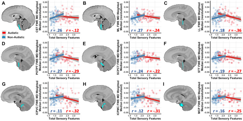
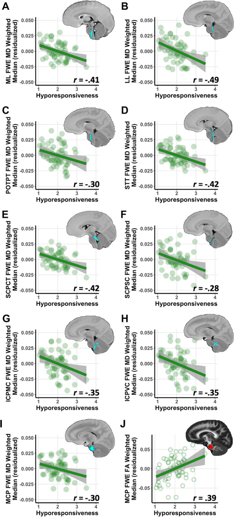
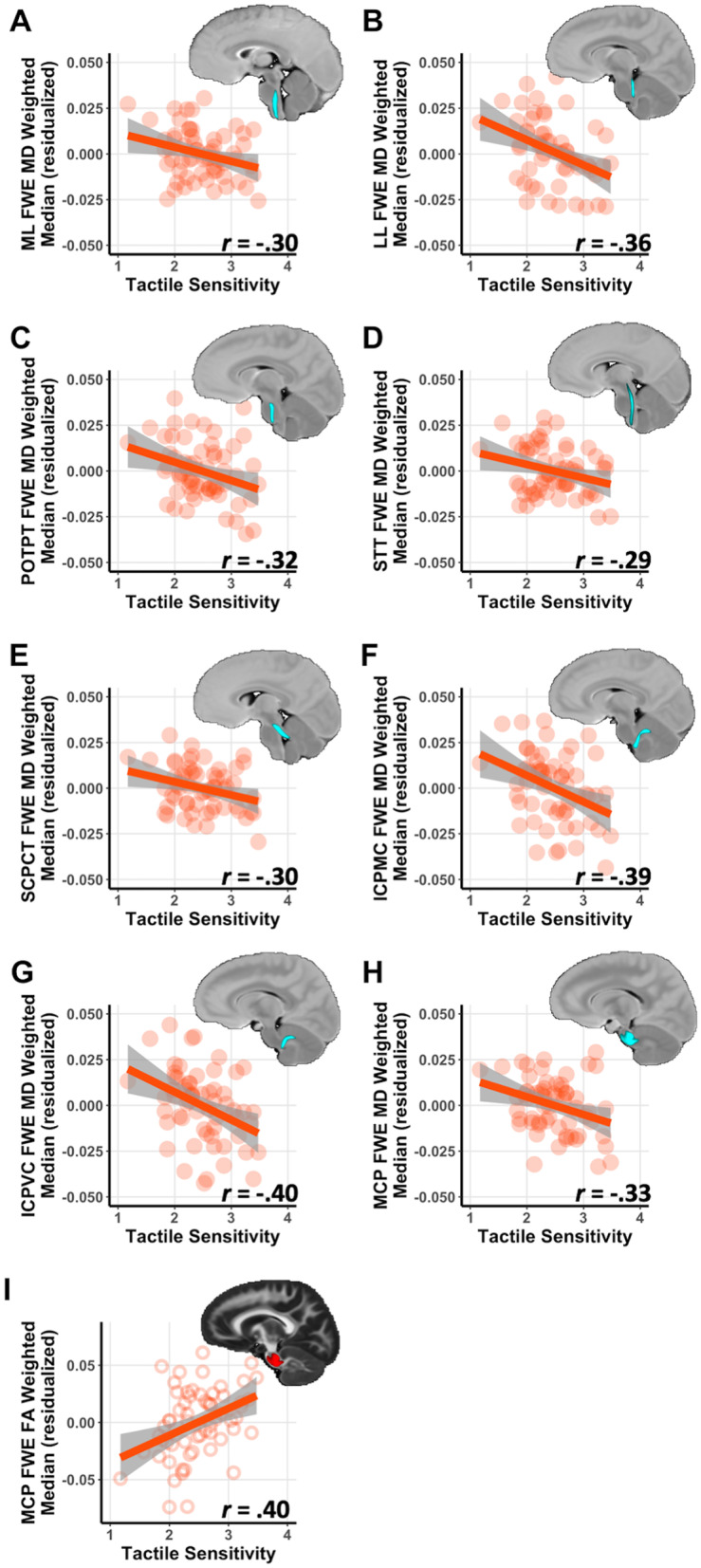
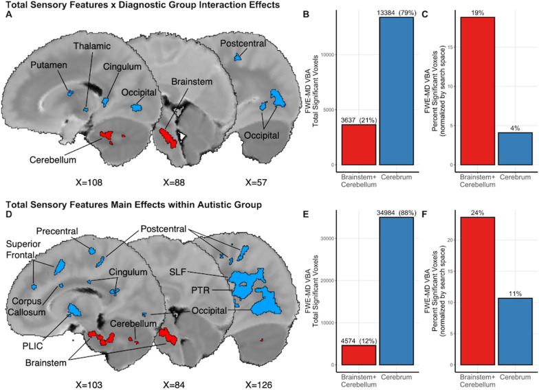
Results: Results revealed distinct relationships between brainstem microstructure and sensory features in autistic children compared to non-autistic children. In autistic children, more prominent sensory features were generally associated with lower MD. Further, in autistic children, sensory hyporesponsiveness and tactile responsivity were strongly associated with white matter microstructure in nearly all brainstem tracts. Follow-up voxel-based analyses confirmed that these relationships were more prominent in the brainstem/cerebellum, with additional sensory-brain findings in the autistic group in the white matter of the primary motor and somatosensory cortices, the occipital lobe, the inferior parietal lobe, and the thalamic projections.
Limitations: All participants communicated via spoken language and acclimated to the sensory environment of an MRI session, which should be considered when assessing the generalizability of this work to the whole of the autism spectrum.
Conclusions: These findings suggest unique brainstem white matter contributions to sensory features in autistic children compared to non-autistic children. The brainstem correlates of sensory features underscore the potential reflex-like nature of behavioral responses to sensory stimuli in autism and have implications for how we conceptualize and address sensory features in autistic populations.
Keywords: Autism; Brainstem; DTI; Sensory features; Voxel-based analysis; White matter.
© 2022. The Author(s).
Conflict of interest statement
ALA is part owner of ImgGyd, LLC and inseRT MRI, Inc. (also listed as TherVoyant). While both companies are involved in developing MRI-based surgery techniques, neither are associated with any current areas of his research, including the present publication. All other authors report no biomedical financial interests of potential conflicts of interest.
Figures
Similar articles
-
Short-Term Memory Impairment.2024 Jun 8. In: StatPearls [Internet]. Treasure Island (FL): StatPearls Publishing; 2025 Jan–. 2024 Jun 8. In: StatPearls [Internet]. Treasure Island (FL): StatPearls Publishing; 2025 Jan–. PMID: 31424720 Free Books & Documents.
-
"It Was Like the Final Piece in the Puzzle for Me": A Qualitative Study on the Experiences of Autistic Women Initially Diagnosed with Borderline Personality Disorder.Autism Adulthood. 2024 Dec 2;6(4):428-437. doi: 10.1089/aut.2023.0031. eCollection 2024 Dec. Autism Adulthood. 2024. PMID: 40018060
-
White matter microstructure of children with sensory over-responsivity is associated with affective behavior.J Neurodev Disord. 2024 Jan 2;16(1):1. doi: 10.1186/s11689-023-09513-w. J Neurodev Disord. 2024. PMID: 38166648 Free PMC article.
-
The Lived Experience of Autistic Adults in Employment: A Systematic Search and Synthesis.Autism Adulthood. 2024 Dec 2;6(4):495-509. doi: 10.1089/aut.2022.0114. eCollection 2024 Dec. Autism Adulthood. 2024. PMID: 40018061 Review.
-
Memantine for autism spectrum disorder.Cochrane Database Syst Rev. 2022 Aug 25;8(8):CD013845. doi: 10.1002/14651858.CD013845.pub2. Cochrane Database Syst Rev. 2022. PMID: 36006807 Free PMC article.
Cited by
-
Cortico-basal ganglia white matter microstructure is linked to restricted repetitive behavior in autism spectrum disorder.Mol Autism. 2024 Jan 23;15(1):6. doi: 10.1186/s13229-023-00581-2. Mol Autism. 2024. PMID: 38254158 Free PMC article.
-
Sex Differences in the Striatal Contributions to Longitudinal Fine Motor Development in Autistic Children.Biol Psychiatry. 2025 Jun 15;97(12):1150-1162. doi: 10.1016/j.biopsych.2025.01.005. Epub 2025 Jan 14. Biol Psychiatry. 2025. PMID: 39818327
-
A multidimensional investigation of the relationship between skin-mediated somatosensory signals, emotion regulation and behavior problems in autistic children.Front Neurosci. 2023 Aug 10;17:1227173. doi: 10.3389/fnins.2023.1227173. eCollection 2023. Front Neurosci. 2023. PMID: 37662109 Free PMC article.
-
Role of autonomic, nociceptive, and limbic brainstem nuclei in core autism features.Autism Res. 2024 Feb;17(2):266-279. doi: 10.1002/aur.3096. Epub 2024 Jan 26. Autism Res. 2024. PMID: 38278763 Free PMC article.
-
Microstructural neural correlates of maximal grip strength in autistic children: the role of the cortico-cerebellar network and attention-deficit/hyperactivity disorder features.Front Integr Neurosci. 2024 May 14;18:1359099. doi: 10.3389/fnint.2024.1359099. eCollection 2024. Front Integr Neurosci. 2024. PMID: 38808069 Free PMC article.
References
Publication types
MeSH terms
Grants and funding
- R00 MH110596/MH/NIMH NIH HHS/United States
- R01 NS117568/NS/NINDS NIH HHS/United States
- R01 NS111022/NS/NINDS NIH HHS/United States
- P01 AI132132/AI/NIAID NIH HHS/United States
- T32 NS105602/NS/NINDS NIH HHS/United States
- R01 NS105646/NS/NINDS NIH HHS/United States
- P50 HD105353/HD/NICHD NIH HHS/United States
- T32 NS076067/NS/NINDS NIH HHS/United States
- U54 HD090256/HD/NICHD NIH HHS/United States
- R01 HD094715/HD/NICHD NIH HHS/United States
- R01 AG037639/AG/NIA NIH HHS/United States
- P30 HD003352/HD/NICHD NIH HHS/United States
- R01 AI138647/AI/NIAID NIH HHS/United States
- T32 CA009206/CA/NCI NIH HHS/United States
LinkOut - more resources
Full Text Sources
