

doi: 10.1002/ctm2.1139.
Raman spectroscopic cellomics for the detection of SARS-CoV-2-associated neutrophil activation after TNF-α stimulation
Affiliations
- PMID: 36536489
- PMCID: PMC9763540
- DOI: 10.1002/ctm2.1139
Item in Clipboard
Raman spectroscopic cellomics for the detection of SARS-CoV-2-associated neutrophil activation after TNF-α stimulation
Clin Transl Med.
2022 Dec.
No abstract available
Conflict of interest statement
The authors declare that they have no conflict of interest.
Figures
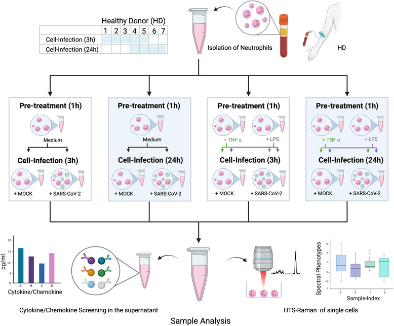
Study flow chart indicating isolation, pre‐treatment, infection and sample analysis. Blood was withdrawn from healthy donors (HD) numbers 1–5 for experiments with 3 h neutrophil‐virus interaction and from HD numbers 4–7 for experiments with 3‐h neutrophil‐virus interaction. In a first step, neutrophils were investigated without any activation by typical chemo‐ or cytokines (pre‐treatment = medium). In the second part, cells were pre‐treated before SARS‐CoV‐2 infection with the specific neutrophil chemoattractant tumor necrosis factor‐alpha (TNF‐α) or lipopolysaccharides (LPS) for 1 h each. Mock and SARS‐CoV‐2 infection were performed independently for each pre‐treatment (indicated by the green and purple arrows in case of pre‐treatment with TNF‐α and LPS in Figure 1). Raman spectra from all datasets collected. Technological specifications of the HTS‐Raman set up are published previously (3, 4). Figure was created with BioRender.com
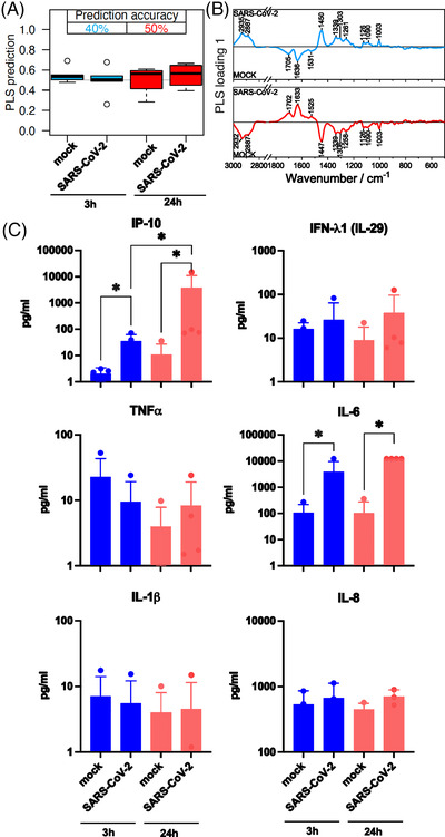
Comparison of neutrophil granulocytes after 3 and 24 h mock and SARS‐CoV‐2 infection. (A) The box plot summarizes the prediction results of the partial least‐squares discriminant analysis (PLS‐DA) model mock versus SARS‐CoV‐2 based on the Raman spectral phenotypes of the cells. Samples with values below 0.5 were predicted as non‐infected cells (not challenged by the virus), and samples with values above 0.5 were predicted as infected cells. Blue boxes reveal the results after a 3‐h infection period; the balanced accuracy of the model is highlighted in blue. Red boxes summarize the results of the 24 h model, and the balanced accuracy is highlighted in red. (B) Loading of the PLS model provide insights which Raman spectral features in the model revealed minor differences between the mock and SARS‐CoV‐2 classes. (C) Bar plots present the mean of all individual values and show the indicated cytokine and chemokine levels measured in the cell supernatant 3 h (blue bars) and 24 h (red bars) p.i. (post infection). Statistical significance was determined via two‐tailed Mann–Whitney test, *p < 0.05. The data that support the findings of this study are available from the corresponding author upon reasonable request.
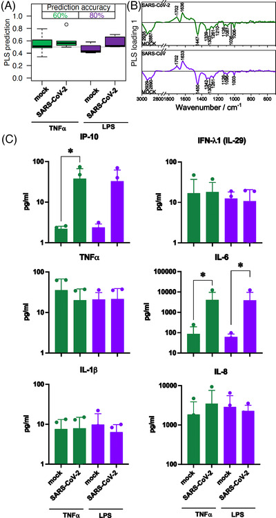
Comparison of neutrophil granulocytes after 1‐h pre‐treatment with TNF‐α and lipopolysaccharides (LPS) followed by a 3‐h mock or SARS‐CoV‐2 infection. (A) The box plot summarizes the prediction results of the model mock versus SARS‐CoV‐2 based on the Raman spectral phenotypes of the cells. Green boxes reveal the results after a TNF‐α pre‐treatment and 3‐h infection period; the balanced accuracy of the model is highlighted in green. Purple boxes summarize the results of the LPS pre‐treatment and 3‐h infection period model, and the balanced accuracy is highlighted in purple. (B) Loading of the partial least‐squares (PLS) model provide insights which Raman spectral features in the model revealed differences between the mock and SARS‐CoV‐2 classes. (C) Bar plots present the mean of all individual values and show the indicated cytokine and chemokine levels measured in the cell supernatant after TNF‐α pre‐treatment (green bars) and LPS pre‐treatment (green bars) and 3‐of infection. Statistical significance was determined via two‐tailed Mann Whitney test *p < 0.05. The data that support the findings of this study are available from the corresponding author upon reasonable request. TNF‐α, tumor necrosis factor‐alpha.
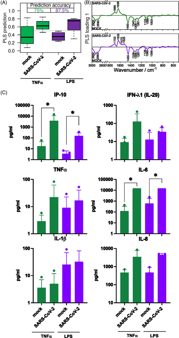
Comparison of neutrophil granulocytes after 1 h pre‐treatment with TNF‐α and lipopolysaccharides (LPS) followed by a 24‐h mock or SARS‐CoV‐2 infection. (A) The box plot summarizes the prediction results of the model mock versus SARS‐CoV‐2 based on the Raman spectral phenotypes of the cells. Green boxes reveal the results after a TNF‐α pre‐treatment and 24‐h infection period, the balanced accuracy of the model is highlighted in green. Purple boxes summarize the results of the LPS pre‐treatment and 24‐h infection period model, and the balanced accuracy is highlighted in purple. (B) Loading of the partial least‐squares (PLS) model provide insights which Raman spectral features in the model revealed differences between the mock and SARS‐CoV‐2 classes. (C) Bar plots present the mean of all individual values and show the indicated cytokine and chemokine levels measured in the cell supernatant after TNF‐α pre‐treatment (green bars) and LPS pre‐treatment (green bars) and 24‐h infection. Statistical significance was determined via two‐tailed Mann–Whitney test *p < 0.05. The data that support the findings of this study are available from the corresponding author upon reasonable request.
References
-
- Schie IW, Ruger J, Mondol AS, et al. High‐throughput screening Raman spectroscopy platform for label‐free cellomics. Anal Chem. 2018;90:2023‐2030. - PubMed
-
- Arend N, Pittner A, Ramoji A, et al. Detection and differentiation of bacterial and fungal infection of neutrophils from peripheral blood using Raman spectroscopy. Anal Chem. 2020;92:10560‐10568. - PubMed
Publication types
MeSH terms
Substances
LinkOut - more resources
Full Text Sources
Medical
Miscellaneous