. 2022 Dec;12(12):e1148.
doi: 10.1002/ctm2.1148.

Artemisitene suppresses rheumatoid arthritis progression via modulating METTL3-mediated N6-methyladenosine modification of ICAM2 mRNA in fibroblast-like synoviocytes

Affiliations

Artemisitene suppresses rheumatoid arthritis progression via modulating METTL3-mediated N6-methyladenosine modification of ICAM2 mRNA in fibroblast-like synoviocytes

Jian Chen et al. Clin Transl Med. 2022 Dec.

Abstract

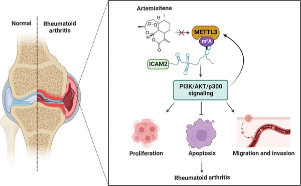
Background: Rheumatoid arthritis (RA) is a chronic autoimmune disease. We previously revealed that the natural compound artemisitene (ATT) exhibits excellent broad anticancer activities without toxicity on normal tissues. Nevertheless, the effect of ATT on RA is undiscovered. Herein, we aim to study the effect and potential mechanism of ATT on RA management.

Methods: A collagen-induced arthritis (CIA) mouse model was employed to confirm the anti-RA potential of ATT. Cell Counting Kit-8 (CCK-8) and 5-ethynyl-2'-deoxyuridine (EdU) assays, cell cycle and apoptosis analysis, immunofluorescence, migration and invasion assays, quantitative real-time PCR (RT-qPCR), Western blot, RNA-sequencing (RNA-seq) analysis, plasmid construction and lentivirus infection, and methylated RNA immunoprecipitation and chromatin immunoprecipitation assays, were carried out to confirm the effect and potential mechanism of ATT on RA management.

Results: ATT relieved CIA in mice. ATT inhibited proliferation and induced apoptosis of RA-fibroblast-like synoviocytes (FLSs). ATT restrained RA-FLSs migration and invasion via suppressing epithelial-mesenchymal transition. RNA-sequencing analysis and bioinformatics analysis identified intercellular adhesion molecule 2 (ICAM2) as a promoter of RA progression in RA-FLSs. ATT inhibits RA progression by suppressing ICAM2/phosphoinositide 3-kinase (PI3K)/protein kinase B (AKT)/p300 pathway in RA-FLSs. Moreover, ATT inhibited methyltransferase-like 3 (METTL3)-mediated N6-methyladenosine methylation of ICAM2 mRNA in RA-FLSs. Interestingly, p300 directly facilitated METTL3 transcription, which could be restrained by ATT in RA-FLSs. Importantly, METTL3, ICAM2 and p300 expressions in synovium tissues of RA patients were related to clinical characteristics and therapy response.

Conclusions: We provided strong evidence that ATT has therapeutic potential for RA management by suppressing proliferation, migration and invasion, in addition to inducing apoptosis of RA-FLSs through modulating METTL3/ICAM2/PI3K/AKT/p300 feedback loop, supplying the fundamental basis for the clinical application of ATT in RA therapy. Moreover, METTL3, ICAM2 and p300 might serve as biomarkers for the therapy response of RA patients.

Keywords: ICAM2; METTL3; artemisitene; fibroblast-like synoviocytes; rheumatoid arthritis.


Conflict of interest statement

The authors declare they have no conflicts of interest.

Figures

FIGURE 1
FIGURE 1
Artemisitene (ATT) significantly relieves collagen‐induced arthritis (CIA) in mice. Mouse models with CIA were established, and methotrexate (MTX) or ATT treatment was carried out three times per week after the second immunisation for the indicated duration. (A and B) The effect of ATT treatment on the incidence (A) and paw severity (B) of CIA mice were assessed after the second immunization (n = 5). (C–I) The swollen joints (C), safranin O (SO) staining (D and E), haematoxylin and eosin (H&E) staining (F), intra‐articular inflammatory cell area (G), and immunofluorescence of vimentin (H and I) of paw sections are shown after ATT treatment for 17 days. Scale bar of SO staining, H&E staining and immunofluorescence are 25, 125 and 12.5 μm, respectively. * p < .05 and ** p < .01 versus CIA group
FIGURE 2
FIGURE 2
Artemisitene (ATT) inhibits proliferation and induces apoptosis of rheumatoid arthritis‐fibroblast‐like synoviocytes (RA‐FLSs). (A) Synovial tissues were obtained from two patients with RA, and RA‐FLSs were identified by immunofluorescence assay using monoclonal antibody vimentin. Scale bar: 25 μm. (B) RA‐FLSs were stimulated with multiple concentrations (2.5, 5, 7.5, 10 and 15 μM) of ATT for 24 h, then CCK‐8 assay was carried out to measure the cell viability (n = 5). (C–F) RA‐FLSs were stimulated with 5, 10 and 15 μM of ATT for 24 h. Representative photographs of the EdU‐positive cells are shown, and the statistical differences were measured after ATT stimulation using EdU assay (n = 3). Scale bar: 25 μm. (G–N) RA‐FLSs from two RA patients were stimulated with ATT for 24 h. Flow cytometry was adopted to estimate the cell cycle (G–J) and apoptotic cells (K–N) after ATT treatment (n = 3). (O–V) The relative mRNA expression of genes associated with cell cycle after ATT treatment for 24 h in RA‐FLSs was measured by RT‐qPCR (n = 3). (W and X) The relative protein expression of proteins associated with cell cycle and apoptosis after ATT treatment for 24 h in RA‐FLSs was measured by Western blot (n = 3). * p < .05 and ** p < .01 versus control
FIGURE 3
FIGURE 3
Artemisitene (ATT) inhibits migration and invasion by suppressing epithelial–mesenchymal transition (EMT) in rheumatoid arthritis‐fibroblast‐like synoviocytes (RA‐FLSs). RA‐FLSs from two patients with RA were treated with ATT (10 μM) for 24 h. (A–C) Representative photographs of migrant and invasive cells (A) are shown, and the statistical differences were measured after ATT stimulation (B and C) (n = 3). Scale bar: 50 μm. (D and E) The relative protein levels of vimentin, E‐cadherin and N‐cadherin were measured by Western blot (n = 3). (F–I) The representative photographs of vimentin, E‐cadherin and N‐cadherin expression were measured by immunofluorescence assay. Scale bar: 50 μm. ** p < .01 versus control
FIGURE 4
FIGURE 4
RNA‐sequencing (RNA‐seq) data analysis of artemisitene (ATT)‐treated rheumatoid arthritis‐fibroblast‐like synoviocytes (RA‐FLSs). RA‐FLSs were treated with ATT (10 μM) for 24 h, and non‐treated RA‐FLSs were used as normal control. Cells were harvested for mRNA sequencing analysis after treatment. (A) The heatmap shows the differentially expressed gene information between the ATT‐treated group and the control group. (B) Volcano diagram shows the differential expression of genes between ATT‐treated group and control group. (C) Kyoto Encyclopedia of Genes and Genomes (KEGG) enrichment analysis showed the differentially enriched signalling pathways, including PI3K–AKT, RA and DNA replication pathways. (D) Gene Ontology (GO) enrichment analysis showed the differential genes were enriched in biological processes, molecular functions and cellular components, including regulation of cell cycle, growth and phosphorylation
FIGURE 5
FIGURE 5
Bioinformatics analysis recognised the rheumatoid arthritis (RA) development associated genes in fibroblast‐like synoviocytes (FLSs). (A and B) The expression patterns of 29 differentially expressed genes between RA synovial fibroblasts (RASF) and healthy synovial fibroblasts (HSF) in GSE21959 and GSE29746, respectively. (C and D) Volcano plots of differentially expressed genes between RASF and HSF in GSE21959 and GSE29746, respectively. (E–J) Bar plots, bubble plots and chords show the top terms of the enriched Gene Ontology (GO) sets and Kyoto Encyclopedia of Genes and Genomes (KEGG) pathways. (K) The intersection of genes in artemisitene (ATT)‐treated RA‐FLSs (Geneset A) identified in gene set enrichment analysis (GSEA) (Geneset B) and genes correlated with clinical characteristics and treatment response of RA patients (Geneset C)
FIGURE 6
FIGURE 6
Artemisitene (ATT) attenuates intercellular adhesion molecule 2 (ICAM2)/PI3K/AKT/p300 pathway in rheumatoid arthritis‐fibroblast‐like synoviocytes (RA‐FLSs). RA‐FLSs from two patients with RA were stimulated with ATT (10 μM) for 24 h. (A–C) The relative mRNA (A) and protein (B and C) expression of ICAM2 after ATT treatment was measured by RT‐qPCR and Western blot in RA‐FLSs (n = 3). (D–F) RT‐qPCR (D) and Western blot (E and F) showing the overexpression efficiency of ICAM2 in RA‐FLSs. (G) RA‐FLSs with or without ICAM2 overexpression (OE‐ICAM2) were stimulated with ATT, then CCK‐8 assay was carried out to measure the cell viability (n = 5). (H–L) Representative photographs showing the apoptotic (H and I), migrant and invasive cells (J–L) after ATT stimulation in RA‐FLSs with or without OE‐ICAM2 (n = 3). Scale bar: 40 μm. (M and N) RA‐FLSs from two patients with RA were stimulated with ATT (10 μM) for 24 h. Western blot showing the relative levels of PI3K, p‐PI3K, AKT, p‐AKT and p300. (O–Q) RT‐qPCR (O) and Western blot (P and Q) showing the knockdown efficiency of ICAM2 in RA‐FLSs, and the relative levels of PI3K, p‐PI3K, AKT, p‐AKT and p300 after knockdown of ICAM2 in RA‐FLSs were measured by Western blot (P and Q) (n = 3). (R and S) The relative levels of PI3K, p‐PI3K, AKT, p‐AKT and p300 after ATT treatment with or without 740Y‐P in RA‐FLSs were measured by Western blot (n = 3). (T) RA‐FLSs with or without 740Y‐P were stimulated with ATT, then CCK‐8 assay was carried out to measure the cell viability (n = 5). (U–Y) Representative photographs showing the apoptotic (U and V), migrant and invasive cells (W–Y) after ATT stimulation in RA‐FLSs with or without 740Y‐P treatment (n = 3). Scale bar: 40 μm. ** p < .01 versus control or negative control (NC); ## p < .01 versus NC + ATT or ATT
FIGURE 7
FIGURE 7
Artemisitene (ATT) inhibits METTL3‐mediated m6A methylation of intercellular adhesion molecule 2 (ICAM2) mRNA and modulates METTL3/ICAM2/PI3K/AKT/p300 feedback loop in rheumatoid arthritis‐fibroblast‐like synoviocytes (RA‐FLSs). (A–C) RA‐FLSs from two patients with RA were treated with ATT (10 μM) for 24 h. RT‐qPCR and Western blot showing the relative mRNA (A) and protein (B and C) expression of METTL3 after ATT treatment in RA‐FLSs (n = 3). (D–F) RT‐qPCR (D) and Western blot (E and F) showing the overexpression (OE) efficiency of METTL3 in RA‐FLSs. (G) RA‐FLSs with or without OE of METTL3 were stimulated with ATT for 24 h, then CCK‐8 assay was carried out to measure the cell viability (n = 5). (H–L) Representative photographs showing the apoptotic (H and I), migrant and invasive cells (J–L) after ATT stimulation in RA‐FLSs with or without METTL3 OE (n = 3). Scale bar: 40 μm. (M–O) RT‐qPCR (M) and Western blot (N and O) showing the knockdown efficiency of METTL3 in RA‐FLSs, and Western blot (N and O) showing the protein expression of ICAM2 after knockdown of METTL3 in RA‐FLSs (n = 3). (P) The m6A modification sites in the 3′‐untranslated region (UTR) of ICAM2 mRNA were revealed through m6A‐REF‐seq and miCLIP in the RMVar and m6A‐Atlas databases. (Q and R) RNA immunoprecipitation (RIP) with anti‐m6A antibody showing the m6A modification of ICAM2 in RA‐FLSs by RT‐PCR (Q) and RT‐qPCR (R). (S and T) RIP with anti‐m6A antibody showing the m6A modification of ICAM2 in RA‐FLSs by RT‐PCR (S) and RT‐qPCR (T). (U–W) RT‐qPCR (U) and Western blot (V and W) showing the knockdown efficiency of p300 in RA‐FLSs (U–W), and Western blot (V and W) showing the protein expression of METTL3 after knockdown of p300 in RA‐FLSs (n = 3). (X) Chromatin immunoprecipitation sequencing (ChIP‐seq) data obtained from Cistrome Data Browser (http://cistrome.org/db/#/; CistromeDB: 36878, CistromeDB: 41271 and CistromeDB: 49414) showing that p300 could bind to the promoter regions of METTL3 in fibroblasts. (Y and Z) ChIP assay showing the inhibitory effect of ATT on METTL3 transcription directly regulated by p300 detected by RT‐PCR (Y) and RT‐qPCR (Z). ** p < .01 versus control or negative control (NC); ## p < .01 versus NC + ATT
FIGURE 8
FIGURE 8
Underlying working model of artemisitene (ATT) in treating rheumatoid arthritis (RA). ATT regulates proliferation, apoptosis as well as migration and invasion of RA‐fibroblast‐like synoviocytes (RA‐FLSs) by modulating the METTL3/intercellular adhesion molecule 2 (ICAM2)/PI3K/AKT/p300 feedback loop, contributing to the suppression of RA progression

Similar articles

Cited by

References

    1. Nygaard G, Firestein GS. Restoring synovial homeostasis in rheumatoid arthritis by targeting fibroblast‐like synoviocytes. Nat Rev Rheumatol. 2020;16(6):316‐333. - PMC - PubMed
    1. Wang Q, Chu P, Yu X, Li J, Zhang W, Gong M. ZFAS1 knockdown inhibits fi broblast‐like synoviocyte proliferation, migration, invasion and inflammation, and promotes apoptosis via miR‐3926/FSTL1 in rheumatoid arthritis. Exp Ther Med. 2021;22(3):914. - PMC - PubMed
    1. Zhang S, Zhao J, Ma W. Circ‐Sirt1 inhibits proliferation, induces apoptosis, and ameliorates inflammation in human rheumatoid arthritis fibroblast‐like synoviocytes. Autoimmunity. 2021;54(8):514‐525. - PubMed
    1. Zhang X, Nan H, Guo J, Liu J. NLRP12 reduces proliferation and inflammation of rheumatoid arthritis fibroblast‐like synoviocytes by regulating the NF‐kappaB and MAPK pathways. Eur Cytokine Netw. 2021; 32(2):15‐22. - PubMed
    1. Mueller AL, Payandeh Z, Mohammadkhani N, et al. Recent advances in understanding the pathogenesis of rheumatoid arthritis: new treatment strategies. Cells. 2021;10(11):3017. - PMC - PubMed

Publication types

MeSH terms