. 2023 Feb:48:101729.
doi: 10.1016/j.nantod.2022.101729. Epub 2022 Dec 15.

SARS-CoV-2 multi-variant rapid detector based on graphene transistor functionalized with an engineered dimeric ACE2 receptor

Affiliations

SARS-CoV-2 multi-variant rapid detector based on graphene transistor functionalized with an engineered dimeric ACE2 receptor

Alice Romagnoli et al. Nano Today. 2023 Feb.

Abstract

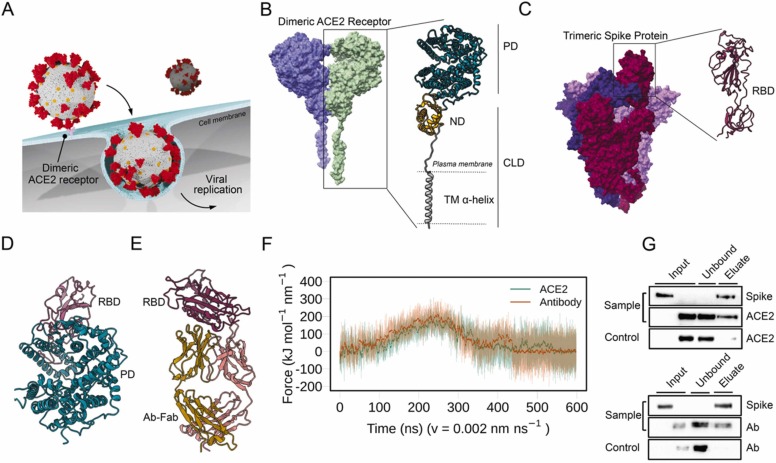
Reliable point-of-care (POC) rapid tests are crucial to detect infection and contain the spread of Severe Acute Respiratory Syndrome Coronavirus 2 (SARS-CoV-2). The emergence of several variants of concern (VOC) can reduce binding affinity to diagnostic antibodies, limiting the efficacy of the currently adopted tests, while showing unaltered or increased affinity for the host receptor, angiotensin converting enzyme 2 (ACE2). We present a graphene field-effect transistor (gFET) biosensor design, which exploits the Spike-ACE2 interaction, the crucial step for SARS-CoV-2 infection. Extensive computational analyses show that a chimeric ACE2-Fragment crystallizable (ACE2-Fc) construct mimics the native receptor dimeric conformation. ACE2-Fc functionalized gFET allows in vitro detection of the trimeric Spike protein, outperforming functionalization with a diagnostic antibody or with the soluble ACE2 portion, resulting in a sensitivity of 20 pg/mL. Our miniaturized POC biosensor successfully detects B.1.610 (pre-VOC), Alpha, Beta, Gamma, Delta, Omicron (i.e., BA.1, BA.2, BA.4, BA.5, BA.2.75 and BQ.1) variants in isolated viruses and patient's clinical nasopharyngeal swabs. The biosensor reached a Limit Of Detection (LOD) of 65 cps/mL in swab specimens of Omicron BA.5. Our approach paves the way for a new and reusable class of highly sensitive, rapid and variant-robust SARS-CoV-2 detection systems.

Keywords: Biosensor; Centaurus; Cerberus; Molecular dynamics; Omicron; Point-of-care; SARS-CoV-2 variants; gFET.


Conflict of interest statement

The authors declare that they have no known competing financial interests or personal relationships that could have appeared to influence the work reported in this paper. The ACE2-Fc functionalization of gFET and the design of the POC device are under patent pending, applied by Polytechnic University of Marche. D.DM., M.DA., C.A., A.R., I.DA., D.M., E.P., P.C., G.B., L.P., M.F. are the inventors of the patent application N. 102021000000533 filed in 01/13/2021. All other authors declare they have no competing interests.

Figures

ga1
We present a graphene field-effect transistor (gFET) biosensor design, which exploits the Spike-ACE2 interaction, essential for infection. Extensive computational analyses show that a chimeric ACE2-Fc construct mimics the native receptor dimer. ACE2-Fc functionalized gFET allows in vitro detection of trimeric Spike, with a limit of detection (LOD) of 20 pg/mL. Our miniaturized POC biosensor successfully detects all prominent virus variants in both isolated viruses and patient’s clinical swabs with a LOD of 65 cps/mL.
Fig. 1
Fig. 1
Probing the interactions of ACE2 and Antibodies with SARS-CoV-2 Spike. A) Schematic representation of ACE2-mediated host cell entry mechanism. B) Cryo-EM structure of dimeric ACE2 receptor (the two monomers are colored differently). The two subunits of a monomer are reported: Peptidase domain (PD) and Collectrin-like domain (CLD) that is composed of neck domain (ND) and transmembrane α-helix (TM α-helix). C) Cryo-EM structure of soluble trimeric Spike protein (the three monomers have different colors). A zoomed-in view of the RBD is shown. D) Peptidase Domain of the ACE2 receptor bound to the RBD of the SARS-CoV-2 Spike protein. E) Antibody Anti-Spike CR3022 (Ab-CR3022), bound to the SARS-CoV-2 Spike protein RBD. F) Force profiles from the Steered Molecular Dynamics (SMD) simulations of RBD unbinding from the ACE2 receptor (orange) and Ab-CR3022 (green). G) Pull-down assay of Spike and ACE2 (upper), and Spike and Ab-CR3022 (lower). Control is represented by the same experiment excluding the Spike protein (bait) from the system. The binding of Spike with ACE2 or Ab-CR3022 were monitored by western blot analysis.
Fig. 2
Fig. 2
gFET setup and Spike recognition. A) gFET (size 10 mm × 10 mm) is composed of two source electrodes each one connected with six graphene channels and the respective drains. A single gate electrode is used for both sides of gFET. A schematic representation of the PBASE-modified gFET is reported in the inset panel. B) Raman Spectra of gFET (black) and gFET-PBASE (red) (diode laser wavelength 523 nm and laser power 50 mW). C) Schematic representation of gFET modified with ACE2. D-E) Detail of Ids-Vg curves obtained for (D) ACE2_gFET (black) and ACE2_gFET + Spike (2 µg/mL) (red). E) ACE2_gFET (black) and ACE2_gFET + mPRO (2 µg/mL) (red). F) Comparative bar charts of ACE2_gFET before (black bars) and after (red bars) the addition of Spike (2 and 0.2 µg/mL) or mPRO (2 µg/mL). G) Schematic representation of gFET modified with Ab (Ab-CR3022). H-I) Detail of Ids-Vg curves for (H) Ab_gFET (black) and Ab_gFET + Spike (2 µg/mL) (red); (I) Ab_gFET (black) and Ab_gFET + mPRO (2 µg/mL) (red); (J) Comparative bar charts of Ab_gFET before (black bars) and after (red bars) the addition of Spike (2, 0.2 and 0.02 µg/mL) or mPRO (2 µg/mL). Details of Vg from 0 to 1.20 V are shown. In (F) and (J): * ** p < 0.001, * p < 0.05, error bars represent standard deviation (s.d.).
Fig. 3
Fig. 3
MD simulations of full length, soluble and Fc tagged ACE2 dimers. A) Representative structure of the full-length ACE2 dimer embedded in a membrane. B) Same as (A) but the membrane is omitted to show the TM helices. C) Soluble truncated ACE2 conformation. D) Starting configuration of the ACE2-Fc chimera. For B-D two orthogonal views are shown. E) Representative snapshots of ACE2-Fc structures sampled during the MD trajectory, side view. The PD centers of mass distance is shown by a dashed red line. F) Top view of soluble ACE2, the PD centers of mass distance is shown by a dashed black line. G) Number of contacts between the two PD-CLD regions of monomers for membrane embedded full length ACE2 (in black), ACE2-Fc (red) and soluble ACE2 (grey). H) Time evolution of the intermonomer distance measured between the PD domains, color code as in (G). Comparative Dynamics as reported by the cross-correlation matrices of concerted motions of the residues of our three dimeric systems during the MD simulations. In (I) correlations in the full length ACE2 embedded in the membrane (upper triangle) and soluble truncated ACE2 (lower triangle). L) Same comparison as (I) between full length ACE2 embedded in the membrane (upper triangle) and ACE2-Fc (lower triangle).
Fig. 4
Fig. 4
ACE2-Fc as bioreceptor and LOD calculation. A) SDS-PAGE under reducing (w β lanes) and non-reducing (w/o β lanes) conditions of soluble ACE2 and ACE2-Fc; B) Pull-down assay of Spike and ACE2-Fc. The binding of Spike with ACE2-Fc was monitored by western blot. C) Schematic representation of gFET modified with ACE2-Fc. D) Comparative bar chart showing the ACE2-Fc_gFET response to different concentrations of SARS-CoV-2 Spike. E) Comparative bar chart showing the ACE2-Fc_gFET response to MERS-CoV Spike protein (2 µg/mL) and SARS-CoV-2 mPRO (2 µg/mL). In (D) and (E): * ** p < 0.001, * * p < 0.01 and * p < 0.05, error bars represent s.d.
Fig. 5
Fig. 5
POC device detects SARS-CoV-2 variants in clinical samples up to low viral concentrations. A) Detailed amino acid mutations of SARS-CoV-2 Spike proteins in B.1.610, Alpha, Delta, Omicron (BA.1, BA.2, BA.4, BA.5 and BQ.1) variants, compared to the wild-type Whuan-Hu1 of SARS-CoV-2. Positions of mutations are shown both on a schematic domain representation of the protein and on the 3D model (PDB ID: 7DWZ). B) Photograph of the gFET Cartridge Unit and the Signal acquisition modules connected to form the entire POC. A reference dimension bar is reported. Also, a schematic representation of gFET modified with ACE2-Fc tested with different SARS-CoV-2 samples is shown. C) Bar graph reporting ACE2-Fc_gFET signal before (black) and after the addition of nasopharyngeal swab samples from patients (red). D) Comparative bar chart showing the ACE2-Fc_gFET response to different dilutions of Omicron BA.5 swab. Ct and cps/mL of each patient or dilution are shown in Tables 1 or 2, respectively. In (C) and (D): ***p < 0.001, **p < 0.01 and * p < 0.05, error bars represent s.d.

References

    1. Grubaugh N.D., Hodcroft E.B., Fauver J.R., Phelan A.L., Cevik M. Cell. 2021;184:1127–1132. doi: 10.1016/j.cell.2021.01.044. - DOI - PMC - PubMed
    1. (2021). https://outbreak.info/ (accessed September 17, 2021).

    1. Barlow P., van Schalkwyk M.C., McKee M., Labonté R., Stuckler D. Lancet planet. Heal. 2021;5:e102–e107. doi: 10.1016/S2542-5196(20)30291-6. - DOI - PMC - PubMed
    1. Zhang Y., Huang Z., Zhu J., Li C., Fang Z., Chen K., Zhang Y. Bioeng. Transl. Med. 2022 doi: 10.1002/btm2.10356. - DOI - PMC - PubMed
    1. Toropov N., Osborne E., Joshi L.T., Davidson J., Morgan C., Page J., Pepperell J., Vollmer F. ACS Sens. 2021;6:2815–2837. doi: 10.1021/acssensors.1c00612. - DOI - PubMed

LinkOut - more resources