

doi: 10.18240/ijo.2022.12.23.
eCollection 2022.
Differentiation of premacular hemorrhages with niveau formation
Affiliations
- PMID: 36536970
- PMCID: PMC9729079
- DOI: 10.18240/ijo.2022.12.23
Item in Clipboard
Differentiation of premacular hemorrhages with niveau formation
Int J Ophthalmol.
.
No abstract available
Figures

A: Fundus photograph shows an intrapocket hemorrhage with niveau formation. There is no spreading of hemorrhage to surrounding tissue (*), except for the hemorrhage in the connecting channel leading to Cloquet's canal (arrow). B: On OCT, no retinal compression (arrow) and the thin anterior wall of the vitreous pocket (arrowhead) can be seen. The hemorrhage is denser in the inferior portion due to gravity. C: The hemorrhage is displaced inferiorly. D: OCT confirms posterior vitreous detachment. Hemorrhage is identified in the vitreous (*) but not in posterior vitreous cortex. OCT: Optical coherence tomography.
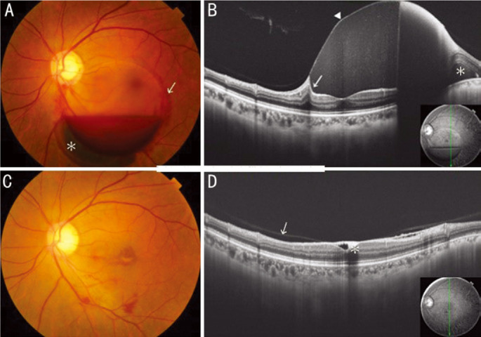
A: Fundus photograph shows a sub-ILM hemorrhage with niveau formation and subretinal hemorrhage (*). Spreading of hemorrhage to surrounding tissue is observed (arrow). B: On OCT, the hemorrhage compresses the retina (arrow) and has uniform density because of the ILM (arrowhead). Subretinal hemorrhage (*) is confirmed. C: Hemorrhage was absorbed after 3mo. D: On OCT, cells depositing on sub-ILM space (*) at the fovea and partial posterior vitreous detachment (arrow) can be seen. OCT: Optical coherence tomography, ILM: Internal limiting membrane.

A: Fundus photograph shows a premacular retrocortical hemorrhage with niveau formation. Bleeding spreads nasally beyond the optic disc, accompanied by vitreous hemorrhage. Spreading of hemorrhage to surrounding tissue is observed (*). B: On OCT, the hemorrhage does not compress the retina (arrow), and posterior vitreous (arrowhead) can be seen. C: Fundus photograph at 6mo after vitrectomy. D: OCT shows no macular edema. OCT: Optical coherence tomography.
References
-
- Celik Dulger S, Ozdal PC, Teke MY. Valsalva retinopathy: long-term results and management strategies. Eur J Ophthalmol. 2021;31(4):1953–1960. - PubMed
-
- Babu N, Kohli P, Rajan RP, Ramasamy K. Inverse drainage Nd:YAG membranotomy for pre-macular hemorrhage. Eur J Ophthalmol. 2022:112067212211022. - PubMed
LinkOut - more resources
Full Text Sources