

A practical approach for PD-L1 evaluation in gastroesophageal cancer
- PMID: 36537078
- PMCID: PMC10462995
- DOI: 10.32074/1591-951X-836
A practical approach for PD-L1 evaluation in gastroesophageal cancer
Abstract
PD-L1 is an established predictive immunohistochemical biomarker of response to immune checkpoint inhibitors. At present, PD-L1 is routinely assessed on biopsy samples of advanced gastroesophageal cancer patients before initiating first-line treatment. However, PD-L1 is still a suboptimal biomarker, due to changing cut-off values and scoring systems, interobserver and interlaboratory variability.
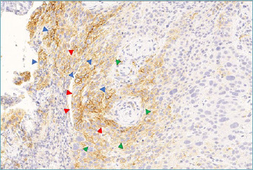
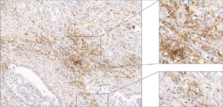
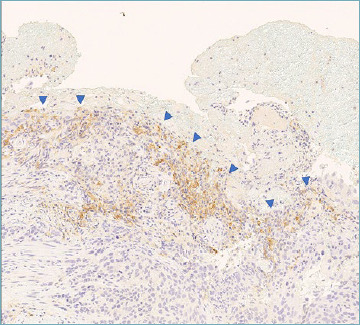
This practical illustrated review discusses the range of staining patterns of PD-L1 and the potential pitfalls and challenges that can be encountered when evaluating PD-L1, focusing on gastric and gastroesophageal adenocarcinoma (G/GEA) and esophageal squamous cell carcinoma (ESCC).
Keywords: PD-L1; esophageal squamous cell carcinoma; gastroesophageal adenocarcinoma; immunohistochemistry; immunotherapy.
Copyright © 2022 Società Italiana di Anatomia Patologica e Citopatologia Diagnostica, Divisione Italiana della International Academy of Pathology.
Conflict of interest statement
The authors declare no conflict of interest related to the present work.
Figures


References
-
- Gou Q, Dong C, Xu H, et al. . PD-L1 degradation pathway and immunotherapy for cancer. Cell Death Dis 2020;11:955. https://doi.org/10.1038/s41419-020-03140-2 10.1038/s41419-020-03140-2 - DOI - PMC - PubMed
-
- Darvin P, Toor SM, Sasidharan Nair V, Elkord E. Immune checkpoint inhibitors: recent progress and potential biomarkers. Exp Mol Med 2018;50:1-11. https://doi.org/10.1038/s12276-018-0191-1 10.1038/s12276-018-0191-1 - DOI - PMC - PubMed
-
- Booth ME, Smyth EC. Immunotherapy in Gastro-Oesophageal Cancer: Current Practice and the Future of Personalised Therapy. BioDrugs 2022;36:473-485. https://doi.org/10.1007/s40259-022-00527-9 10.1007/s40259-022-00527-9 - DOI - PubMed
-
- Mastracci L, Grillo F, Parente P, et al. . PD-L1 evaluation in the gastrointestinal tract: from biological rationale to its clinical application. Pathologica 2022;114:352-364. https://doi.org/10.32074/1591-951X-803 10.32074/1591-951X-803 - DOI - PMC - PubMed
-
- Ajani JA, D’Amico TA, Bentrem DJ, et al. . Gastric Cancer, Version 2.2022, NCCN Clinical Practice Guidelines in Oncology. J Natl Compr Canc Netw 2022;20:167-192. https://doi.org/10.6004/jnccn.2022.0008 10.6004/jnccn.2022.0008 - DOI - PubMed
Publication types
MeSH terms
Substances
LinkOut - more resources
Full Text Sources
Medical
Research Materials
