. 2023 May 2;119(4):969-981.
doi: 10.1093/cvr/cvac189.

Cholesterol accumulation in macrophages drives NETosis in atherosclerotic plaques via IL-1β secretion

Affiliations

Cholesterol accumulation in macrophages drives NETosis in atherosclerotic plaques via IL-1β secretion

Mustafa Yalcinkaya et al. Cardiovasc Res. .

Abstract

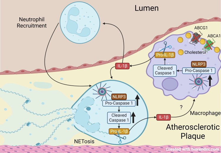
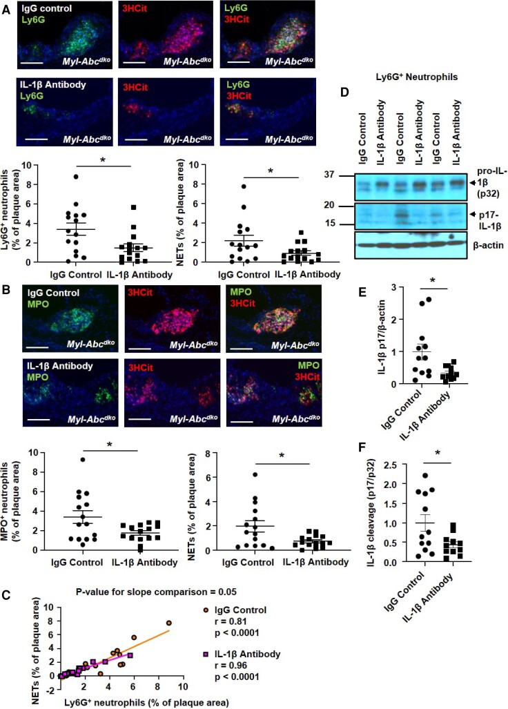
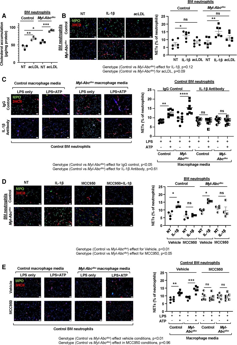
Aims: Neutrophil extracellular trap formation (NETosis) increases atherosclerotic plaque vulnerability and athero-thrombosis. However, mechanisms promoting NETosis during atherogenesis are poorly understood. We have shown that cholesterol accumulation due to myeloid cell deficiency of the cholesterol transporters ATP Binding Cassette A1 and G1 (ABCA1/G1) promotes NLRP3 inflammasome activation in macrophages and neutrophils and induces prominent NETosis in atherosclerotic plaques. We investigated whether NETosis is a cell-intrinsic effect in neutrophils or is mediated indirectly by cellular crosstalk from macrophages to neutrophils involving IL-1β.

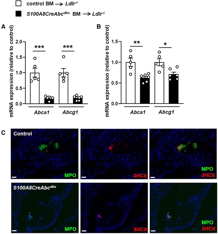
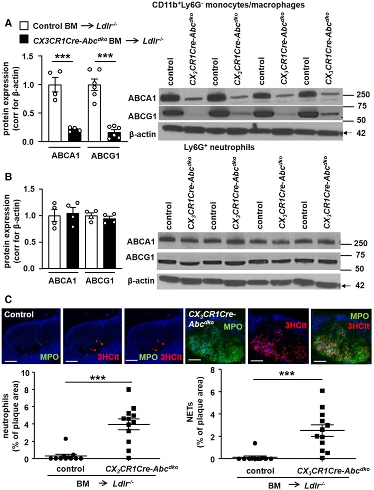
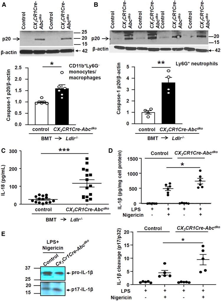
Methods and results: We generated mice with neutrophil or macrophage-specific Abca1/g1 deficiency (S100A8CreAbca1fl/flAbcg1fl/fl or CX3CR1CreAbca1fl/flAbcg1fl/fl mice, respectively), and transplanted their bone marrow into low-density lipoprotein receptor knockout mice. We then fed the mice a cholesterol-rich diet. Macrophage, but not neutrophil Abca1/g1 deficiency activated inflammasomes in macrophages and neutrophils, reflected by caspase-1 cleavage, and induced NETosis in plaques. NETosis was suppressed by administering an interleukin (IL)-1β neutralizing antibody. The extent of NETosis in plaques correlated strongly with the degree of neutrophil accumulation, irrespective of blood neutrophil counts, and neutrophil accumulation was decreased by IL-1β antagonism. In vitro, IL-1β or media transferred from Abca1/g1-deficient macrophages increased NETosis in both control and Abca1/Abcg1 deficient neutrophils. This cell-extrinsic effect of IL-1β on NETosis was blocked by an NLRP3 inhibitor.

Conclusion: These studies establish a new link between inflammasome-mediated IL-1β production in macrophages and NETosis in atherosclerotic plaques. Macrophage-derived IL-1β appears to increase NETosis both by increasing neutrophil recruitment to plaques and by promoting neutrophil NLRP3 inflammasome activation.

Keywords: Atherosclerosis; Inflammation; Leukocyte.


Conflict of interest statement

A.R.T. is a consultant for Amgen, CSL Behring, Astra Zeneca, and Foresite Laboratories, and is on the SAB of Staten Biotech, Fortico Biotech, and Beren Therapeutics. P.L. is an unpaid consultant to, or involved in clinical trials for Amgen, AstraZeneca, Baim Institute, Beren Therapeutics, Esperion Therapeutics, Genentech, Kancera, Kowa Pharmaceuticals, Medimmune, Merck, Norvo Nordisk, Novartis, Pfizer, and Sanofi-Regeneron. P.L. is a member of the scientific advisory board for Amgen, Caristo Diagnostics, Cartesian Therapeutics, CSL Behring, DalCor Pharmaceuticals, Dewpoint Therapeutics, Euclid Bioimaging, Kancera, Kowa Pharmaceuticals, Olatec Therapeutics, Medimmune, Moderna, Novartis, PlaqueTec, TenSixteen Bio, Soley Therapeutics, and XBiotech, Inc. P.L.’s laboratory has received research funding in the last 2 years from Novartis. P.L. is on the Board of Directors of XBiotech, Inc. P.L. has a financial interest in Xbiotech, a company developing therapeutic human antibodies, in TenSixteen Bio, a company targeting somatic mosaicism and CHIP to discover and develop novel therapeutics to treat age-related diseases, and in Soley Therapeutics, a biotechnology company that is combining artificial intelligence with molecular and cellular response detection for discovering and developing new drugs, currently focusing on cancer therapeutics. P.L.'s interests were reviewed and are managed by Brigham and Women's Hospital and Partners HealthCare in accordance with their conflict-of-interest policies.

Figures

Graphical abstract
Graphical abstract
Figure 1
Figure 1
Neutrophil Abca1/g1 deficiency does not affect NETosis in atherosclerotic plaques of Ldlr−/− mice. Ldlr−/− mice were transplanted with bone marrow (BM) from Abca1fl/flAbcg1fl/fl (control) or S100A8CreAbca1fl/flAbcg1fl/fl (S100A8-Cre-Abcdko) mice and fed Western-type diet (WTD) for 8 weeks. (A–B) Ly6G+ neutrophils (A) and Ly6GCD11b+ monocytes (B) were isolated from BM and Abca1 and Abcg1 mRNA expression was assessed. n = 5. *P < 0.05, by t-test. (C) Mice were sacrificed and hearts were isolated, sectioned, and neutrophils were stained in atherosclerotic lesions of the aortic root using myeloperoxidase (MPO). Lesions were also stained for citrullinated histones 2, 8, and 17 (3HCit). NETs show an overlap of MPO and 3HCit staining. Representative pictures are shown. Scale bars: 25 μm.
Figure 2
Figure 2
Macrophage Abca1/g1 deficiency induces neutrophil accumulation and NETosis in atherosclerotic lesions. Ldlr−/− mice were transplanted with BM from Abca1fl/flAbcg1fl/fl (control) or CX3CR1CreAbca1fl/flAbcg1fl/fl (CX3CR1Cre-Abcdko) mice and fed WTD for 8 weeks. Ly6GCD11b+ splenic monocytes/macrophages (A) and Ly6G+ splenic neutrophils (B) were isolated and Abca1 and Abcg1 protein expression was assessed by Western blot, corrected for β-actin, and quantified. n = 4. Unedited gels are shown in Supplementary material online, Figure S16. (C) In atherosclerotic lesions, neutrophils were stained using MPO, and the MPO+ percentage of lesion size was quantified. Lesions were also stained for 3HCit. To assess NETs, the overlap of MPO and 3HCit was quantified. Representative pictures are shown. Scale bars: 75 μm. Each datapoint represents an individual mouse. n = 12. ***P < 0.001, by t-test.
Figure 3
Figure 3
Macrophage Abca1/g1 deficiency induces inflammasome activation in monocytes, macrophages, and neutrophils of Ldlr−/− mice. Ldlr−/− mice were transplanted with BM from Abca1fl/flAbcg1fl/fl (control) or CX3CR1CreAbca1fl/flAbcg1fl/fl (CX3CR1Cre-Abcdko) mice and fed WTD for 8 weeks. Ly6GCD11b+ splenic monocytes/macrophages (A) and Ly6G+ splenic neutrophils (B) were isolated and caspase-1 cleavage was assessed by Western blot. To quantify caspase-1 cleavage, the p20 cleaved form of caspase-1 was divided by the p45 pro-form. n = 4–6. (C) Plasma IL-18 levels. (D–E) IL-1β secretion from control or CX3CR1Cre-Abcdko BMDMs treated with 20 ng/mL LPS for 3 h, and with 10 µg/mL nigericin for an additional 1 h. (D) IL-1β secretion was assessed by ELISA. (E) Immunoblot of IL-1β cleavage of cell lysates. Unedited gels are shown in Supplementary material online, Figure S17. Each datapoint represents an individual mouse. n = 14. *P < 0.05, ** P < 0.01, ***P < 0.001 (A–C) by t-test and (D–E) by one-way ANOVA.
Figure 4
Figure 4
IL-1β antagonism decreases neutrophil content and NETosis in atherosclerotic lesions of Ldlr−/− mice with myeloid Abca1/g1 deficiency. Ldlr−/− mice were transplanted with BM from LysmCreAbca1fl/flAbcg1fl/fl (Myl-Abcdko) mice and fed WTD. The anti-mouse monoclonal antibodies (IgG2a) that selectively neutralize IL-1β or isotype-matched control IgG2a were administered subcutaneously at 6, 7, and 8 weeks of WTD feeding (three doses of antibodies in total). At 8.5 weeks of WTD, mice were sacrificed and neutrophils were stained in atherosclerotic lesions using Ly6G (A) or MPO (B), and Ly6G+ (A) or MPO+ (B) percentages of lesion size were quantified. Concomitantly, lesions were stained for 3HCit. To assess NETs, the overlap of Ly6G and 3HCit (A) or MPO and 3HCit (B) was quantified as a percentage of the total lesion area. Representative pictures are shown. Scale bars: 100 μm. (C) Correlation between Ly6G+ and Ly6G + 3HCit+ (NET) area as shown in (A). (D) Ly6G+ splenic neutrophils were isolated and the cleaved and pro-form of IL-1β was assessed by Western blot. Unedited gels are shown in Supplementary material online, Figure S18. Cleaved IL-1β was quantified relative to β-actin (E) and the pro-form of IL-1β (F). n = 12. Each data point represents an individual mouse. n = 15. *P < 0.05, by t-test.
Figure 5
Figure 5
Macrophage-derived IL-1β induces neutrophil inflammasome and NETosis. (A–B) Ly6G+ neutrophils from BM of Abca1fl/flAbcg1fl/fl (control) or LysmCreAbca1fl/flAbcg1fl/fl (Myl-Abcdko) mice were treated with 100 ng/ml IL-1β and/or 25 µg/mL acLDL for 3 h. (A) Total cholesterol in Ly6G+ neutrophils. (B) Neutrophils were stained for DAPI, MPO, and 3HCit. To assess NETs in isolated neutrophils, the overlap of 3HCit and MPO was quantified and expressed as % of total cells. (C) Control or Myl-Abcdko BM-derived macrophages (BMDMs) were treated with LPS and ATP to activate the NLRP3 inflammasome. Cells were washed and incubated for 1 h with medium to allow for cytokine secretion. The medium was collected and mixed 1:1 with DMEM supplemented with 1% pen-strep and then transferred to neutrophils that had been pre-treated with 100 µg/mL IgG control or IL-1β antibodies for 30 min and incubated with these cells for 3 h. (D) Ly6G+ neutrophils from BM of control or Myl-Abcdko were treated with MCC950 for 30 min and then 100 ng/ml IL-1β was added for 3 h. (E) Ly6G+ neutrophils were pre-treated with MCC950 for 30 min, and subsequently with conditioned media as described in (C). For (C–E), NETosis was assessed as in (B). In B–E, representative pictures are shown. Scale bars: 25 µm. ****P < 0.0001, ***P < 0.001, ** P < 0.01, *P < 0.05, by one-way ANOVA with Tukey’s multiple comparison test (A) or by two-way ANOVA with Sidak’s multiple comparison test (B–E). NT, not treated. Genotype effects per condition are indicated for panels B–E.

References

    1. Vandanmagsar B, Youm YH, Ravussin A, Galgani JE, Stadler K, Mynatt RL, Ravussin E, Stephens JM, Dixit VD. The NLRP3 inflammasome instigates obesity-induced inflammation and insulin resistance. Nat Med 2011;17:179–188. - PMC - PubMed
    1. Duewell P, Kono H, Rayner KJ, Sirois CM, Vladimer G, Bauernfeind FG, Abela GS, Franchi L, Nunez G, Schnurr M, Espevik T, Lien E, Fitzgerald KA, Rock KL, Moore KJ, Wright SD, Hornung V, Latz E. NLRP3 Inflammasomes are required for atherogenesis and activated by cholesterol crystals. Nature 2010;464:1357–1361. - PMC - PubMed
    1. Ridker PM, Everett BM, Thuren T, MacFadyen JG, Chang WH, Ballantyne C, Fonseca F, Nicolau J, Koenig W, Anker SD, Kastelein JJP, Cornel JH, Pais P, Pella D, Genest J, Cifkova R, Lorenzatti A, Forster T, Kobalava Z, Vida-Simiti L, Flather M, Shimokawa H, Ogawa H, Dellborg M, Rossi PRF, Troquay RPT, Libby P, Glynn RJ, Group CT. Antiinflammatory therapy with canakinumab for atherosclerotic disease. N Engl J Med 2017;377:1119–1131. - PubMed
    1. Brinkmann V, Reichard U, Goosmann C, Fauler B, Uhlemann Y, Weiss DS, Weinrauch Y, Zychlinsky A. Neutrophil extracellular traps kill bacteria. Science 2004;303:1532–1535. - PubMed
    1. Borissoff JI, Joosen IA, Versteylen MO, Brill A, Fuchs TA, Savchenko AS, Gallant M, Martinod K, Ten Cate H, Hofstra L, Crijns HJ, Wagner DD, Kietselaer B. Elevated levels of circulating DNA and chromatin are independently associated with severe coronary atherosclerosis and a prothrombotic state. Arterioscler Thromb Vasc Biol 2013;33:2032–2040. - PMC - PubMed

Publication types

MeSH terms

Substances