Clinical Trial
. 2023 Apr;307(1):e212779.
doi: 10.1148/radiol.212779. Epub 2022 Dec 20.

Radiographic Cortical Thickness Index Predicts Fragility Fracture in Gaucher Disease

Collaborators, Affiliations
Clinical Trial

Radiographic Cortical Thickness Index Predicts Fragility Fracture in Gaucher Disease

Simona D'Amore et al. Radiology. 2023 Apr.

Abstract

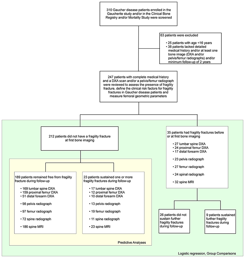
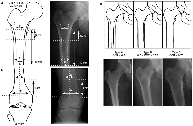
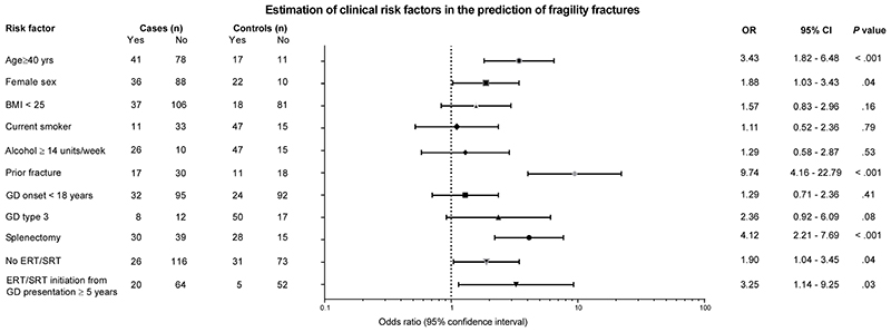
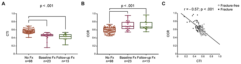
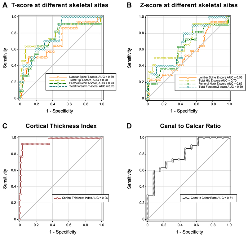
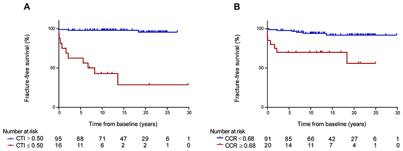
Background Patients with Gaucher disease (GD) have a high risk of fragility fractures. Routine evaluation of bone involvement in these patients includes radiography and repeated dual-energy x-ray absorptiometry (DXA). However, osteonecrosis and bone fracture may affect the accuracy of DXA. Purpose To assess the utility of DXA and radiographic femoral cortical thickness measurements as predictors of fragility fracture in patients with GD with long-term follow-up (up to 30 years). Materials and Methods Patients with GD age 16 years and older with a detailed medical history, at least one bone image (DXA and/or radiographs), and minimum 2 years follow-up were retrospectively identified using three merged UK-based registries (Gaucherite study, enrollment 2015-2017; Clinical Bone Registry, enrollment 2003-2006; and Mortality Registry, enrollment 1993-2019). Cortical thickness index (CTI) and canal-to-calcar ratio (CCR) were measured by two independent observers, and inter- and intraobserver reliability was calculated. The fracture-predictive value of DXA, CTI, CCR, and cutoff values were calculated using receiver operating characteristic curves. Statistical differences were assessed using univariable and multivariable analysis. Results Bone imaging in 247 patients (123 men, 124 women; baseline median age, 39 years; IQR, 27-50 years) was reviewed. The median follow-up period was 11 years (IQR, 7-19 years; range, 2-30 years). Thirty-five patients had fractures before or at first bone imaging, 23 patients had fractures after first bone imaging, and 189 patients remained fracture-free. Inter- and intraobserver reproducibility for CTI/CCR measurements was substantial (range, 0.96-0.98). In the 212 patients with no baseline fracture, CTI (cutoff, ≤0.50) predicted future fractures with higher sensitivity and specificity (area under the receiver operating characteristic curve [AUC], 0.96; 95% CI: 0.84, 0.99; sensitivity, 92%; specificity, 96%) than DXA T-score at total hip (AUC, 0.78; 95% CI: 0.51, 0.91; sensitivity, 64%; specificity, 93%), femoral neck (AUC, 0.73; 95% CI: 0.50, 0.86; sensitivity, 64%; specificity, 73%), lumbar spine (AUC, 0.69; 95% CI: 0.49, 0.82; sensitivity, 57%; specificity, 63%), and forearm (AUC, 0.78; 95% CI: 0.59, 0.89; sensitivity, 70%; specificity, 70%). Conclusion Radiographic cortical thickness index of 0.50 or less was a reliable independent predictor of fracture risk in Gaucher disease. Clinical trial registration no. NCT03240653 © RSNA, 2022 Supplemental material is available for this article.


Conflict of interest statement

Declaration of interests

SD reports speaker fees from Takeda and travel support from Sanofi Genzyme and Takeda outside of the submitted work; and has been recently employed by Chiesi (after the submission of this manuscript). PD reports research grant support from Sanofi Genzyme and Takeda; speaker fee from Sanofi Genzyme and Takeda outside of the submitted work; and is a member of European Board of the International Collaborative Gaucher Group Gaucher Registry (ICGG), which is sponsored by Sanofi Genzyme, and of the Gaucher Outcome Survey Board Member, which is sponsored by Takeda. TMC reports research grant support from Sanofi and Takeda; consulting and speaker fees from Avrobio, Sanofi Genzyme, and Takeda; travel support from Sanofi Genzyme; and advisory board fees from Sanofi Genzyme outside of the submitted work. UR reports research grant support from Amicus, Intrabio, and Takeda; consulting and speaker fees, and travel support from Amicus, Sanofi Genzyme, and Takeda outside of the submitted work. All other authors declare no competing interests.

Figures

Figure 1
Figure 1. Strobe flow chart shows patient selection
DXA = dual-energy X-ray absorptiometry.
Figure 2
Figure 2. Radiographic measurements
(A) The CTI is defined as the ratio of cortical width (a) minus endosteal width (b) to cortical width at a level of 10 cm below the minor trochanter. The CCR is defined as the ratio between the intramedullary canal isthmus (b) and the calcar isthmus (c). (B) Dorr classification of the proximal femoral morphology: type A (CCR<0.50) is defined by thick cortices producing a narrow and funnel shape diaphyseal canal, type B (0.500.75) is characterized by seriously thin cortices resulting in a wide cylindrical shape femoral canal. (C) The metaphyseal index is defined as the ratio of the width of diametaphysis 4 cm from the physeal plate divided by the physeal plate width. Radiographs from (A) a 43-year-old woman with Gaucher disease type 1 and no history of fragility fracture, splenectomy nor osteonecrosis; (B, left panel) and (C) a 29-year-old woman with Gaucher disease type 1 and no history of fragility fracture, splenectomy nor osteonecrosis (right panel); (B, central panel) a 42-year-old woman with Gaucher disease type 1 and prior splenectomy, with no history of fragility fracture nor osteonecrosis; (B, right panel) a 43-year-old woman with Gaucher disease type 1 and history of prior splenectomy, osteonecrosis of the left femoral head, and fragility fracture. CTI=cortical thickness index; CCR=canal to calcar ratio; MI=metaphyseal index.
Figure 3
Figure 3. Clinical risk factors for fractures in patients with Gaucher disease
In the overall study sample (123M:124F, median age 39 years [IQR 27-50]), logistic regression analysis was used to assess the association between fragility fracture and potential risk factors. Horizontal lines indicate 95% confidence interval. BMI=body mass index; CI=confidence interval; ERT=enzyme replacement therapy; GD=Gaucher disease; IQR=interquartile range; OR=odds ratio; SRT=substrate reduction therapy.
Figure 4
Figure 4. Cortical thickness index and canal to calcar ratio in patients with Gaucher disease
Within the whole cohort of 247 patients studied, 134 patients had adequate hip radiographs available for analysis (98 [50M:48F; median age 38 years, IQR 25-48] who remained fracture-free during follow-up; 23 [11M:12F; median age 50 years, IQR 40-56] with fracture before or at first bone imaging; and 13 [4M:9F; median age 40 years, IQR 22-53] who sustained a fracture after first bone imaging). Analysis of differences between groups indicated that patients with GD who sustained fragility fractures before or at first bone imaging (Baseline Fx) or after the first bone imaging (Follow-up Fx) had a smaller (A) CTI (Kruskal-Wallis One-Way ANOVA on Ranks test: P<.001) and larger (B) CCR (Kruskal-Wallis One-Way ANOVA on Ranks test: P<.001) compared with patient who remained free from fracture (No Fx). Boxes include the data between first and third quartiles, the central bar indicates the median and the whiskers show minimum and maximum values. The dots represent all patients. In the overall study sample, the cortical thickness index was negatively correlated to (C) canal to calcar ratio (r=-0.57, P<.001). The correlation between continuous variables was assessed with Spearman’s rank correlation coefficient. (o) Fracture-free; (•) Fracture. CCR=canal to calcar ratio; CTI=cortical thickness index; Fx=fracture; GD=Gaucher disease; IQR=interquartile range.
Figure 5
Figure 5. Performance of dual-energy X-ray absorptiometry, cortical thickness index and canal to calcar ratio as biomarkers of fragility fracture in patients with Gaucher disease
In patients with Gaucher disease with no fracture at baseline, receiver-operating characteristic (ROC) curve analysis was used to assess the performance in the prediction of future fragility fractures of (A) T-score at lumbar spine (n=149 [82M:67F] fracture-free; n=14 [6M:8F] follow-up fracture), total hip (n=147 [82M:65F] fracture-free; n=11 [5M:6F] follow-up fracture), femoral neck (n=144 [80M:64F] fracture-free; n=11 [5M:6F] follow-up fracture), and total forearm (n=50 [21M:29F] fracture-free; n=10 [4M:6F] follow-up fracture); and Z-score at lumbar spine (n=155 [84M:71F] fracture-free; n=17 [7M:10F] follow-up fracture), total hip (n=150 [82M:68F] fracture-free; n=12 [6M:6F] follow-up fracture), femoral neck (n=150 [86M:68F] fracture-free; n=11 [5M:6F] follow-up fracture), and total forearm (n=45 [18M:27F] fracture-free; n=10 [4M:6F] follow-up fracture); and (C and D) CTI and CCR (n=98 [50M:48F] fracture-free during follow-up; n=13 [4M:9F] follow-up fracture). AUC=area under the curve; CCR=canal to calcar ratio; CTI=cortical thickness index; IQR=interquartile range.
Figure 6
Figure 6. Fracture-free survival analysis of cortical thickness index and canal to calcar ratio in patients with Gaucher disease
Within the 212 patients with Gaucher disease who were fracture-free at baseline, 111 patients had adequate hip radiographs available for fracture-free survival analysis (98 [50M:48F; median age 38 years, IQR 25-48] remained fracture-free during follow-up and 13 [4M:9F; median age 40 years, IQR 22-53] who sustained a fracture after first bone imaging). (A) Kaplan-Meier curves of fracture-free survival of patients with Gaucher disease with CTI at baseline below and above 0.50 (log-rank test P<.001). (B) Kaplan-Meier curves of fracture-free survival of patients with Gaucher disease with CCR at baseline below and above 0.68 (log-rank test P<.001). CCR=canal to calcar ratio; CTI=cortical thickness index; IQR=interquartile range.

Comment in

References

    1. Roh J, Subramanian S, Weinreb NJ, Kartha RV. Gaucher disease - more than just a rare lipid storage disease. J Mol Med (Berl) 2022;100:499–518. - PubMed
    1. Hughes D, Mikosch P, Belmatoug N, et al. Gaucher Disease in Bone: From Pathophysiology to Practice. J Bone Miner Res. 2019;34:996–1013. - PMC - PubMed
    1. Wenstrup RJ, Kacena KA, Kaplan P, et al. Effect of enzyme replacement therapy with imiglucerase on BMD in type 1 Gaucher disease. J Bone Miner Res. 2007;22:119–126. - PubMed
    1. Khan A, Hangartner T, Weinreb NJ, Taylor JS, Mistry PK. Risk factors for fractures and avascular osteonecrosis in type 1 Gaucher disease: a study from the International Collaborative Gaucher Group (ICGG) Gaucher Registry. J Bone Miner Res. 2012;27:1839–1848. - PubMed
    1. Leslie WD, Tsang JF, Caetano PA, Lix LM. Effectiveness of bone density measurement for predicting osteoporotic fractures in clinical practice. J Clin Endocrinol Metab. 2007;92:77–81. - PubMed

Publication types

Associated data