. 2022 Dec 20;8(1):149.
doi: 10.1186/s40798-022-00545-5.

Evidence-Based High-Loading Tendon Exercise for 12 Weeks Leads to Increased Tendon Stiffness and Cross-Sectional Area in Achilles Tendinopathy: A Controlled Clinical Trial

Affiliations

Evidence-Based High-Loading Tendon Exercise for 12 Weeks Leads to Increased Tendon Stiffness and Cross-Sectional Area in Achilles Tendinopathy: A Controlled Clinical Trial

Goran Radovanović et al. Sports Med Open. .

Abstract

Background: Assuming that the mechanisms inducing adaptation in healthy tendons yield similar responses in tendinopathic tendons, we hypothesized that a high-loading exercise protocol that increases tendon stiffness and cross-sectional area in male healthy Achilles tendons may also induce comparable beneficial adaptations in male tendinopathic Achilles tendons in addition to improving pain and function.

Objectives: We investigated the effectiveness of high-loading exercise in Achilles tendinopathy in terms of inducing mechanical (tendon stiffness, maximum strain), material (Young's modulus), morphological (tendon cross-sectional area (CSA)), maximum voluntary isometric plantar flexor strength (MVC) as well as clinical adaptations (Victorian Institute of Sports Assessment-Achilles (VISA-A) score and pain (numerical rating scale (NRS))) as the primary outcomes. As secondary outcomes, drop (DJ) and counter-movement jump (CMJ) height and intratendinous vascularity were assessed.

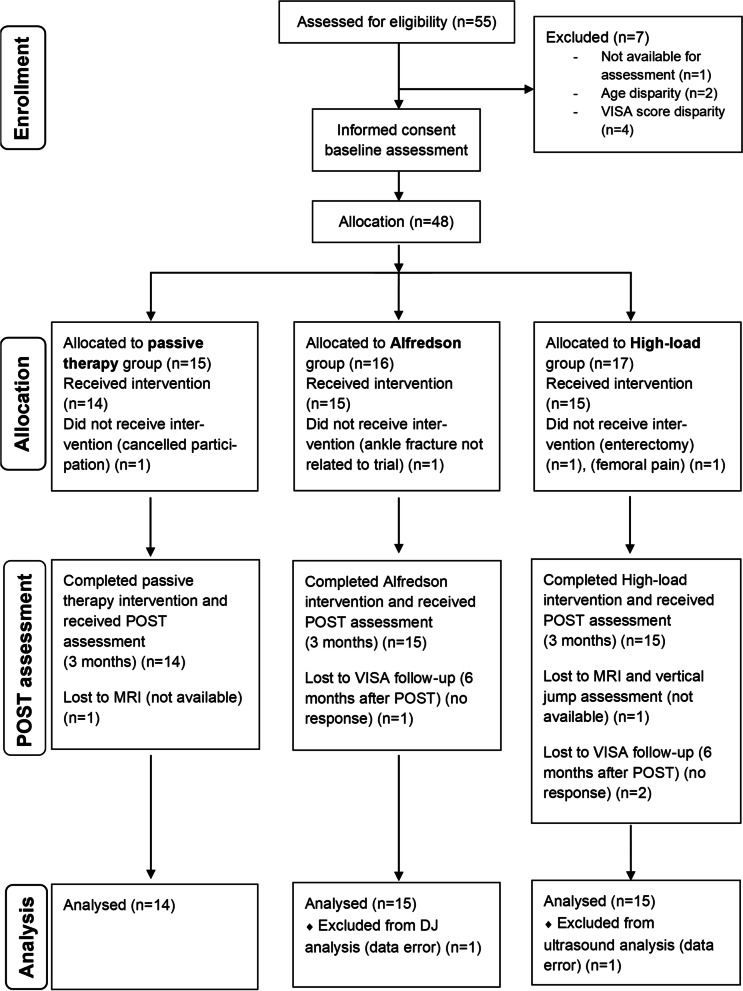
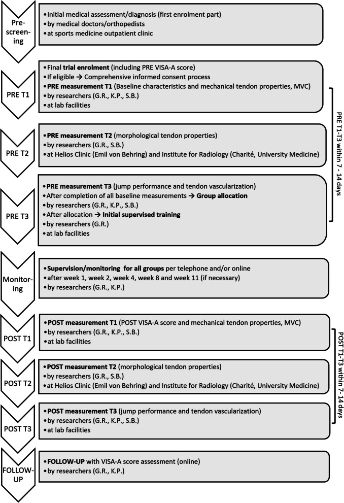
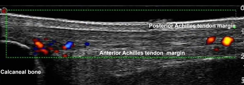
Methods: We conducted a controlled clinical trial with a 3-month intervention phase. Eligibility criteria were assessed by researchers and medical doctors. Inclusion criteria were male sex, aged between 20 and 55 years, chronic Achilles tendinopathy confirmed by a medical doctor via ultrasound-assisted assessment, and a severity level of less than 80 points on the VISA-A score. Thirty-nine patients were assigned by sequential allocation to one of three parallel arms: a high-loading intervention (training at ~ 90% of the MVC) (n = 15), eccentric exercise (according to the Alfredson protocol) as the standard therapy (n = 15) and passive therapy (n = 14). Parameters were assessed pre- and-post-intervention. Data analysis was blinded.

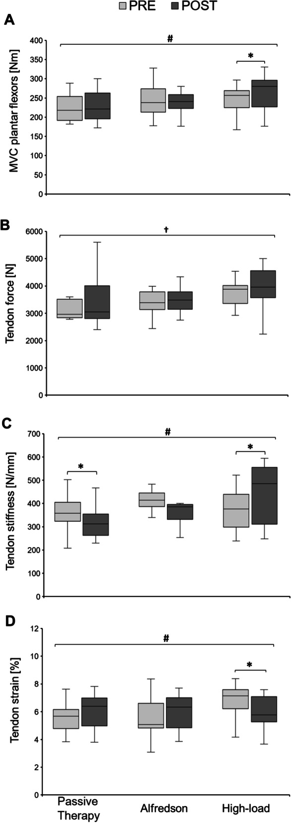
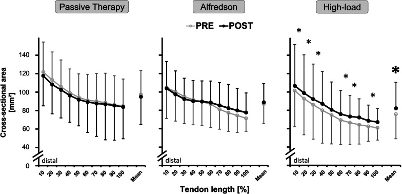
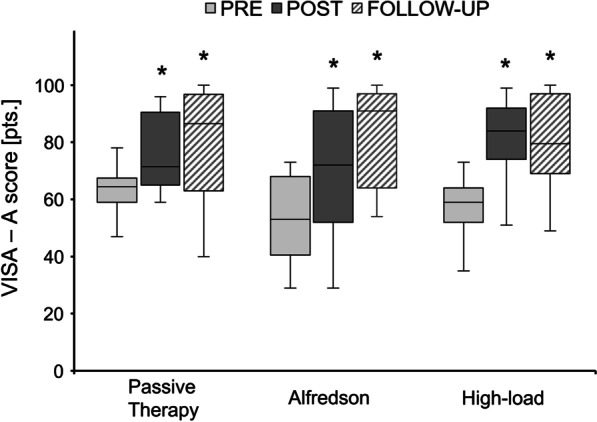
Results: Primary outcomes: Plantar flexor MVC, tendon stiffness, mean CSA and maximum tendon strain improved only in the high-loading intervention group by 7.2 ± 9.9% (p = 0.045), 20.1 ± 20.5% (p = 0.049), 8.98 ± 5.8% (p < 0.001) and -12.4 ± 10.3% (p = 0.001), respectively. Stiffness decreased in the passive therapy group (-7.7 ± 21.2%; p = 0.042). There was no change in Young's modulus in either group (p > 0.05). The VISA-A score increased in all groups on average by 19.8 ± 15.3 points (p < 0.001), while pain (NRS) dropped by -0.55 ± 0.9 points (p < 0.001).

Secondary outcomes: CMJ height decreased for all groups (-0.63 ± 4.07 cm; p = 0.005). There was no change in DJ height and vascularity (p > 0.05) in either group.

Conclusion: Despite an overall clinical improvement, it was exclusively the high-loading intervention that induced significant mechanical and morphological adaptations of the plantar flexor muscle-tendon unit. This might contribute to protecting the tendon from strain-induced injury. Thus, we recommend the high-loading intervention as an effective (alternative) therapeutic protocol in Achilles tendinopathy rehabilitation management in males.

Clinical trials registration number: NCT02732782.

Keywords: Achilles tendinopathy; Controlled clinical trial; Eccentric training; High-loading exercise; Musculoskeletal rehabilitation; Physiotherapy; Tendon adaptation; Tendon rehabilitation; Training therapy.


Conflict of interest statement

The authors declare that they have no competing interests.

Figures

Fig. 1
Fig. 1
Participant flowchart according to the CONSORT (Consolidated Standards of Reporting Trials) guidelines [42]. At baseline and after completion of the 12-week intervention phase, we assessed the mechanical, material, morphological, functional, and clinical properties of the muscle–tendon complex. VISA = Victorian Institute of Sport Assessment questionnaire; MRI = Magnetic Resonance Imaging; DJ = drop jump
Fig. 2
Fig. 2
Data collection timeline flowchart. PRE = before the intervention phase, T = timepoint, VISA-A = Victorian Institute of Sports Assessment—Achilles, POST = after completion of 12 weeks of intervention, FOLLOW-UP = 6 months after completion of the intervention phase
Fig. 3
Fig. 3
Sagittal power Doppler ultrasound scan of the intratendinous vascularity of a tendinopathic Achilles tendon. The green box (indicated by a thin dotted green line) shows the standardized rectangle frame (10.0 × 2.0 cm) of the region of interest in which the Doppler signal was visualized. Within this box, we analyzed vascularity within tendinous tissue only between the anterior margin (i.e., anterior margin of the Achilles tendon and the posterior margin of the calcaneal bone) and posterior margin of the Achilles tendon
Fig. 4
Fig. 4
A–D Muscle strength and tendon mechanical properties of the symptomatic leg at baseline (PRE) and after the 12-week intervention phase (POST) for all three intervention groups. A Isometric maximum voluntary contraction (MVC) of the plantar flexor muscles. B Tendon force of the Achilles tendon. C Tendon stiffness of the Achilles tendon. D Maximum Achilles tendon strain. The horizontal line in the middle of the box is the median value of the scores, and the lower and upper boundaries indicate the 25th and 75th percentiles, respectively (median included). The largest and smallest observed values that are not outliers are shown by the lines drawn from the ends of the box to those values (whiskers). * indicates significant post hoc difference when compared to PRE (p < 0.05); † indicates significantly difference to PRE (p < 0.05) as a main effect of time; # significance with p = 0.05 as a group-by-time interaction effect
Fig. 5
Fig. 5
Cross-sectional area (CSA) values (mm2) of the symptomatic Achilles tendon measured by magnetic resonance imaging (MRI) at baseline (PRE) and after the 12-week intervention phase (POST) for each intervention group (Passive therapy n = 13, Alfredson n = 15, High-load n = 14). Data are presented as CSA values measured in 10% increments tendon length from distal to proximal alongside the free Achilles tendon and the total free Achilles tendon CSA mean ± standard deviation. * indicates significant post hoc difference when compared to the corresponding tendon region in the passive therapy and Alfredson group (p < 0.05). * indicates significant post hoc difference when compared to PRE (p < 0.05)
Fig. 6
Fig. 6
Victorian Institute of Sport Assessment (VISA)—Achilles (A) score at baseline (PRE), after the 12-week intervention period (POST) (Passive therapy n = 14; Alfredson n = 15; High-load n = 15) and 6 months after POST (FOLLOW-UP) (Passive therapy n = 14; Alfredson n = 13; High-load n = 13) for the three intervention groups measured in points (pts.) from 0 to 100. The horizontal line in the middle of the box is the median value of the scores, and the lower and upper boundaries indicate the 25th and 75th percentiles, respectively (median included). The largest and smallest observed values that are not outliers are shown by the lines drawn from the ends of the box to those values (whiskers). * indicates significant difference (p < 0.05) compared to PRE as a significant main effect of time

References

    1. Skjong CC, Meininger AK, Ho SSW. Tendinopathy treatment: where is the evidence? Clin Sports Med. 2012;31:329–350. doi: 10.1016/j.csm.2011.11.003. - DOI - PubMed
    1. de Jonge S, van den Berg C, de Vos RJ, van der Heide HJL, Weir A, Verhaar JAN, et al. Incidence of midportion Achilles tendinopathy in the general population. Br J Sports Med. 2011;45:1026–1028. doi: 10.1136/bjsports-2011-090342. - DOI - PubMed
    1. Andres BM, Murrell GAC. Treatment of tendinopathy: what works, what does not, and what is on the horizon. Clin Orthop Relat Res. 2008;466:1539–1554. doi: 10.1007/s11999-008-0260-1. - DOI - PMC - PubMed
    1. Legerlotz K. Rehabilitation of tendopathies in human athletes. Comp Exerc Physiol. 2013;9:153–160. doi: 10.3920/CEP13030. - DOI
    1. van der Vlist AC, Breda SJ, Oei EHG, Verhaar JAN, de Vos R-J. Clinical risk factors for Achilles tendinopathy: a systematic review. Br J Sports Med. 2019;53:1352–1361. doi: 10.1136/bjsports-2018-099991. - DOI - PMC - PubMed

Associated data