. 2022 Dec 20;17(12):e0276115.
doi: 10.1371/journal.pone.0276115. eCollection 2022.

State-of-the-art analytical methods of viral infections in human lung organoids

Affiliations

State-of-the-art analytical methods of viral infections in human lung organoids

Morris Baumgardt et al. PLoS One. .

Erratum in

Abstract

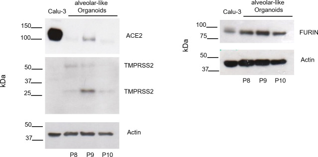
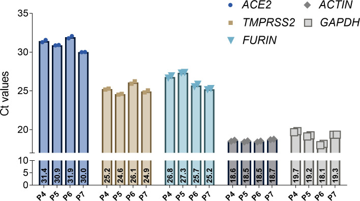
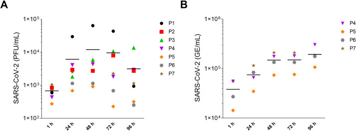
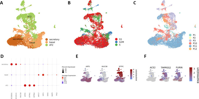
Human-based organ models can provide strong predictive value to investigate the tropism, virulence, and replication kinetics of viral pathogens. Currently, such models have received widespread attention in the study of SARS-CoV-2 causing the COVID-19 pandemic. Applicable to a large set of organoid models and viruses, we provide a step-by-step work instruction for the infection of human alveolar-like organoids with SARS-CoV-2 in this protocol collection. We also prepared a detailed description on state-of-the-art methodologies to assess the infection impact and the analysis of relevant host factors in organoids. This protocol collection consists of five different sets of protocols. Set 1 describes the protein extraction from human alveolar-like organoids and the determination of protein expression of angiotensin-converting enzyme 2 (ACE2), transmembrane serine protease 2 (TMPRSS2) and FURIN as exemplary host factors of SARS-CoV-2. Set 2 provides detailed guidance on the extraction of RNA from human alveolar-like organoids and the subsequent qPCR to quantify the expression level of ACE2, TMPRSS2, and FURIN as host factors of SARS-CoV-2 on the mRNA level. Protocol set 3 contains an in-depth explanation on how to infect human alveolar-like organoids with SARS-CoV-2 and how to quantify the viral replication by plaque assay and viral E gene-based RT-qPCR. Set 4 provides a step-by-step protocol for the isolation of single cells from infected human alveolar-like organoids for further processing in single-cell RNA sequencing or flow cytometry. Set 5 presents a detailed protocol on how to perform the fixation of human alveolar-like organoids and guides through all steps of immunohistochemistry and in situ hybridization to visualize SARS-CoV-2 and its host factors. The infection and all subsequent analytical methods have been successfully validated by biological replications with human alveolar-like organoids based on material from different donors.


Conflict of interest statement

The authors declare no conflict of interest.

Figures

Fig 1
Fig 1. Analysis of SARS-CoV-2 host factor expression by Western blot.
Representative Western blots for ACE2, TMPRSS2, and FURIN of protein lysates from human alveolar-like organoids of three different donors (P8-P10). Calu-3 cells served as positive control.
Fig 2
Fig 2. Analysis of ACE2, TMPRSS2, and FURIN expression in human alveolar-like organoids.
Expression of ACE2, TMPRSS2, FURIN and GAPDH and β-ACTIN as housekeeping genes measured by qPCR on bulk RNA of human alveolar-like organoids. Shown are Ct values of four different donors (P4-P7) and two technical replicates.
Fig 3
Fig 3. Replication kinetics of SARS-CoV-2-infected human alveolar-like organoids.
Human alveolar-like organoids were infected with SARS-CoV-2 (MOI = 1) and viral replication was assessed by plaque assay (A) and via viral E gene-based RT-qPCR (B). Data of seven (plaque assay) and four (RT-qPCR) biological replicates are shown as individual data points. The mean is visualized by a horizontal black line.
Fig 4
Fig 4. Single-cell RNA sequencing displays the proportion of three different cell types and SARS-CoV-2 host factors present in uninfected human alveolar-like organoids.
UMAP embedding of human alveolar-like organoids (n = 6; P1-P3 and P10-P12) shows AT2, basal, and secretory cells present in the organoids (A). Cell cycle phase of each cell in either G2M, S or G1 phase (B) and individual donor composition (C) is displayed. The marker genes needed for cell type identification are shown (D and E) as well as the expression of the SARS-CoV-2 host factors ACE2, TMPRSS2, and FURIN (F).
Fig 5
Fig 5. Spectral imaging of mock- and SARS-CoV-2-infected and immunostained human alveolar-like organoids and in situ hybridization.
(A) Representative immunostainings for exemplary mock- (left panel) and SARS-CoV-2-infected (right column) human alveolar-like organoids. Shown are immunostainings for SARS-CoV-2 (N-protein, red), ACE2 (green) and via in situ hybridization visualized ACE2 mRNA expression (red dots) 24 h post infection (MOI = 1). Arrows indicate either cells positive for SARS-CoV-2 (red arrows) or areas of particularly high ACE2 expression (protein: green arrows, mRNA: red dotted arrows). Cell nuclei are visualized by DAPI stain (blue). Scale bars = 20 μm. (B) In situ hybridization of human alveolar-like organoids shows SARS-CoV-2 mRNA expression (left column). DapB and EF1a served as negative respectively positive control. Red arrows indicate cells positive for SARS-CoV-2. Scale bars = 20 μm.

References

    1. Adhikary PP, Ul Ain Q, Hocke AC, Hedtrich S. COVID-19 highlights the model dilemma in biomedical research. Nat Rev Mater. 2021;6(5):374–6. doi: 10.1038/s41578-021-00305-z - DOI - PMC - PubMed
    1. Kang I, Smirnova L, Kuhn JH, Hogberg HT, Kleinstreuer NC, Hartung T. COVID-19—prime time for microphysiological systems, as illustrated for the brain. ALTEX. 2021;38(4):535–49. doi: 10.14573/altex.2110131 - DOI - PubMed
    1. Munoz-Fontela C, Widerspick L, Albrecht RA, Beer M, Carroll MW, de Wit E, et al.. Advances and gaps in SARS-CoV-2 infection models. PLoS Pathog. 2022;18(1):e1010161. doi: 10.1371/journal.ppat.1010161 - DOI - PMC - PubMed
    1. Hoffmann K, Obermayer B, Honzke K, Fatykhova D, Demir Z, Lowa A, et al.. Human alveolar progenitors generate dual lineage bronchioalveolar organoids. Commun Biol. 2022;5(1):875. doi: 10.1038/s42003-022-03828-5 - DOI - PMC - PubMed
    1. Clevers H. Modeling Development and Disease with Organoids. Cell. 2016;165(7):1586–97. doi: 10.1016/j.cell.2016.05.082 - DOI - PubMed

Publication types