Review
. 2023 Jun;21(6):347-360.
doi: 10.1038/s41579-022-00833-7. Epub 2022 Dec 20.

Microbiota-mediated colonization resistance: mechanisms and regulation

Affiliations
Review

Microbiota-mediated colonization resistance: mechanisms and regulation

Gustavo Caballero-Flores et al. Nat Rev Microbiol. 2023 Jun.

Abstract

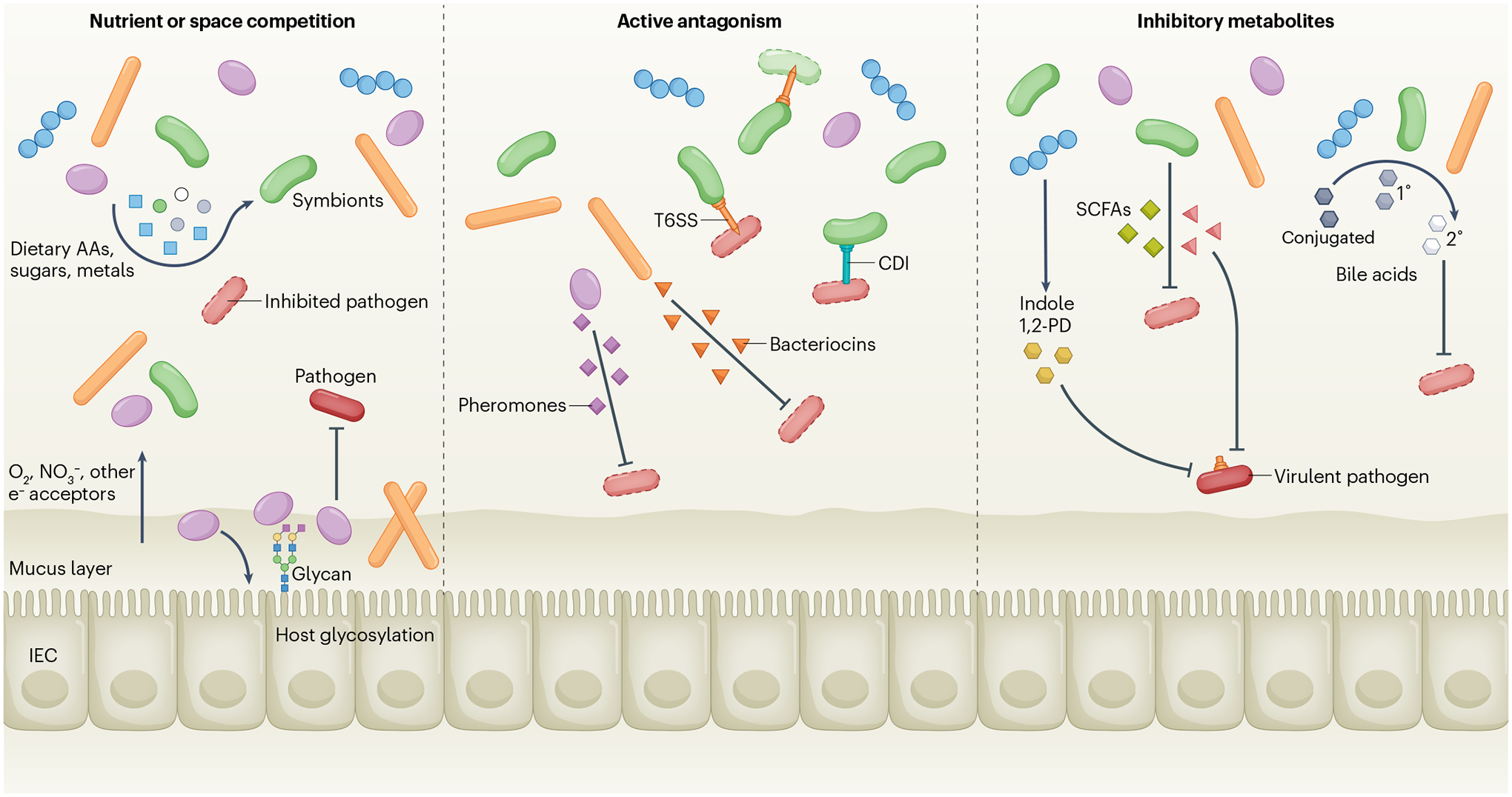
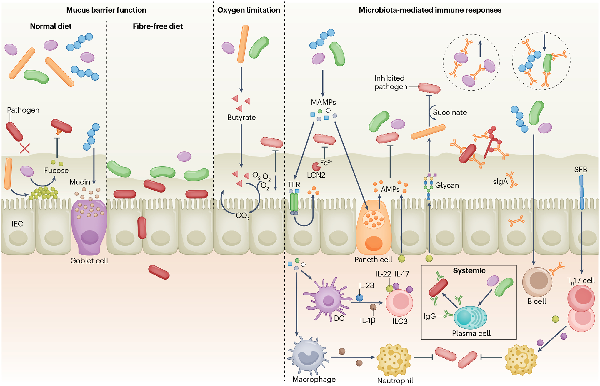
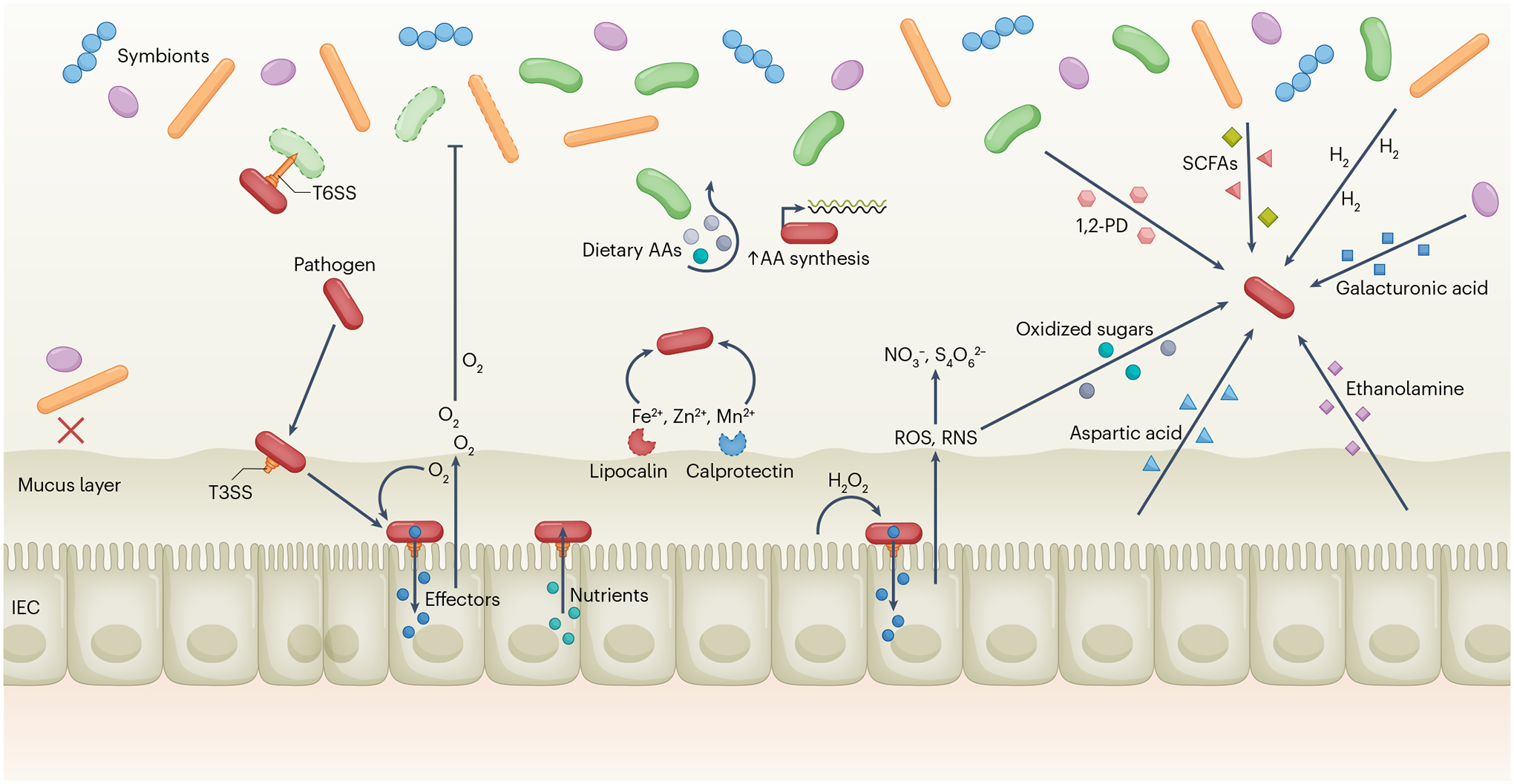
A dense and diverse microbial community inhabits the gut and many epithelial surfaces. Referred to as the microbiota, it co-evolved with the host and is beneficial for many host physiological processes. A major function of these symbiotic microorganisms is protection against pathogen colonization and overgrowth of indigenous pathobionts. Dysbiosis of the normal microbial community increases the risk of pathogen infection and overgrowth of harmful pathobionts. The protective mechanisms conferred by the microbiota are complex and include competitive microbial-microbial interactions and induction of host immune responses. Pathogens, in turn, have evolved multiple strategies to subvert colonization resistance conferred by the microbiota. Understanding the mechanisms by which microbial symbionts limit pathogen colonization should guide the development of new therapeutic approaches to prevent or treat disease.


Conflict of interest statement

Competing interests

The authors declare no competing interests.

Figures

Fig. 1 |
Fig. 1 |. Direct mechanisms of colonization resistance.
Native symbiotic bacteria consume dietary amino acids (AAs), sugars, metals and respiratory electron acceptors such as O2 and NO3, thus starving the pathogen of essential nutrients and molecules (left panel). At the epithelial surface, bacteria can modify host glycosylation and/or use it as a nutrient for adhesion, creating a new microscopic niche that blocks pathogen access to the epithelium. Other symbionts adhering to sites on the epithelium or in the mucus can also prevent pathogen access. Symbionts can also directly kill pathogens via contact-dependent inhibition (CDI), the type VI secretion system (T6SS) or secreted molecules, including bacteriocins or pheromone peptides (centre panel). Other inhibitory compounds such as acetic, butyric or propionic acids (short-chain fatty acids (SCFAs)), indole or 1,2-propanediol (1,2-PD) are secreted by the microbiota and inhibit growth or suppress virulence factor expression in pathogens (right panel). Many symbionts can modify bile acids by deconjugation, whereas some (for example, Clostridium scindens) can convert primary to secondary bile acids that inhibit pathogen growth. IEC, intestinal epithelial cell.
Fig. 2 |
Fig. 2 |. Indirect mechanisms of colonization resistance.
The mucus layer limits pathogen access to the intestinal epithelium (left panel). Microbiota stimulation of host fucosylation protects against pathogens, whereas certain symbionts (such as Bacteroides thetaiotaomicron) release l-fucose from the mucus layer, which reduces virulence gene expression in Citrobacter rodentium, pathogenic Escherichia coli and Enterococcus faecalis. Fibre deprivation leads to an increase in mucus-degrading symbionts that erode the mucus layer, promoting C. rodentium early access to the epithelium. Clostridia produce butyrate that promotes aerobic respiration in intestinal epithelial cells (IECs), reducing oxygen levels in the gut and limiting the expansion of facultative anaerobe pathogens (centre panel). Microorganism-associated molecular patterns (MAMPs) stimulate Toll-like receptors (TLRs) and NOD-like receptors (not shown) in IECs and Paneth cells, leading to the production of antimicrobial peptides (AMPs) that target symbiotic bacteria and pathogens (right panel). Likewise, the microbiota promotes lipocalin 2 (LCN2) production in IECs, limiting pathogen access to iron. Symbiotic bacteria stimulate IL-22 production by type 3 innate lymphoid cells (ILC3s), which regulates AMP production and glycosylation-dependent expansion of symbionts that compete with Clostridioides difficile for succinate. Symbionts also promote pro-IL-1β production by resident macrophages, priming these cells to produce IL-1β in response to Salmonella enterica subsp. enterica serovar Typhimurium and enhancing the recruitment of neutrophils to fight infection. Symbiotic bacteria induce production of polyreactive secretory IgA (sIgA) that can recognize antigens on enteric pathogens, including S. Typhimurium, pathogenic E. coli and virulent Candida albicans. sIgA also limits or promotes expansion of specific symbionts. Microbiota-induced IgG confers protection against pathogens primarily at systemic sites. Segmented filamentous bacteria (SFB) induce IL-17 and IL-22 production by T helper 17 (TH17) cells and ILC3s, promoting neutrophil recruitment and pathogen control. DC, dendritic cell.
Fig. 3 |
Fig. 3 |. Pathogen evasion of colonization resistance.
Upon entering the gut, some pathogens can use a type VI secretion system (T6SS) to directly kill competitors and open a niche (upper left). Most symbionts and pathobionts are excluded from the inner mucus layer (lower left). Certain pathogens, such as Citrobacter rodentium, utilize a type III secretion system (T3SS) to adhere to the epithelium and inject effectors into intestinal epithelial cells (IECs) to modify their physiology, including hyperproliferation of epithelial cells, which causes O2 to be released and respired by the pathogen while killing oxygen-sensitive symbiotic bacteria. C. rodentium can also respire hydrogen peroxide (H2O2) produced by IECs during its early, T3SS-mediated access to the epithelium. Enteropathogenic Escherichia coli can also use its T3SS to obtain nutrients directly from the host cell cytoplasm. Dietary amino acids (AAs) are depleted by the microbiota, but pathogens, including C. rodentium, can synthesize AAs de novo to circumvent this nutrient deficiency (centre). Salmonella enterica subsp. enterica serovar Typhimurium has evolved resistant siderophores and higher-affinity transporters to overcome lipocalin 2-mediated and calprotectin-mediated metal (Fe2+, Zn2+, Mn2+) sequestration. Injection of effectors by the T3SS also causes host inflammatory responses, releasing reactive oxygen species (ROS) and reactive nitrogen species (RNS), which produce respiratory electron acceptors such as tetrathionate (S4O62−) and nitrate (NO3) that can fuel pathogen growth. Oxidized sugars (for example, glucarate and galactarate) are also produced by RNS and used by pathogens. Aspartic acid is released during inflammation and can be utilized for energy production by pathogens. In turn, 1,2-propanediol (1,2-PD), propionic and butyric acids (short-chain fatty acids (SCFAs)), H2 and galacturonic acid are produced by the microbiota and can be metabolized by certain pathogens, as can host-derived ethanolamine, if the appropriate electron acceptors are present.

References

    1. Peterson J et al. The NIH human microbiome project. Genome Res 19, 2317–2323 (2009). - PMC - PubMed
    1. Gu S et al. Bacterial community mapping of the mouse gastrointestinal tract. PLoS ONE 8, e74957 (2013). - PMC - PubMed
    1. Nava GM, Friedrichsen HJ & Stappenbeck TS Spatial organization of intestinal microbiota in the mouse ascending colon. ISME J 5, 627–638 (2011). - PMC - PubMed
    1. Clemente JC, Ursell LK, Parfrey LW & Knight R The impact of the gut microbiota on human health: an integrative view. Cell 148, 1258–1270 (2012). - PMC - PubMed
    1. Kamada N, Chen GY, Inohara N & Núñez G Control of pathogens and pathobionts by the gut microbiota. Nat. Immunol 14, 685–690 (2013). - PMC - PubMed

Publication types