. 2022 Dec 20;12(1):205.
doi: 10.1186/s13578-022-00943-y.

Phosphorylation of adducin-1 by TPX2 promotes interpolar microtubule homeostasis and precise chromosome segregation in mouse oocytes

Affiliations

Phosphorylation of adducin-1 by TPX2 promotes interpolar microtubule homeostasis and precise chromosome segregation in mouse oocytes

Ying Zhang et al. Cell Biosci. .

Abstract

Background: ADD1 (adducin-1) and TPX2 (targeting protein for Xklp2) are centrosomal proteins and regulate mitotic spindle assembly. Mammalian oocytes that segregate homologous chromosomes in Meiosis I and sister chromatids in Meiosis II with a spindle lacking centrosomes are more prone to chromosome segregation errors than in mitosis. However, the regulatory mechanisms of oocyte spindle assembly and the functions of ADD1 and TPX2 in this process remain elusive.

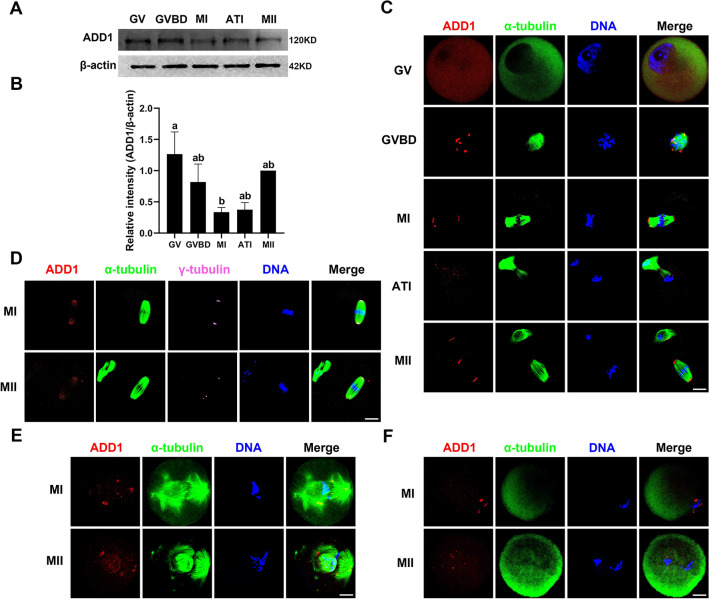
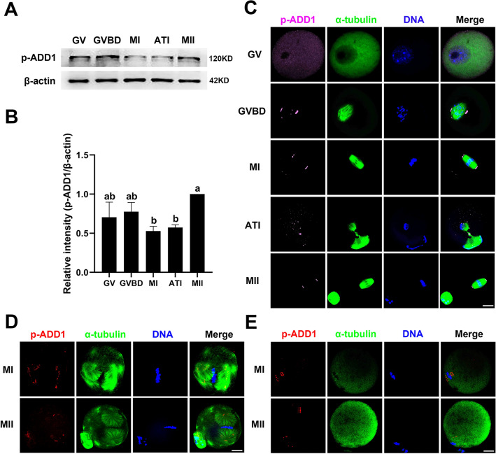
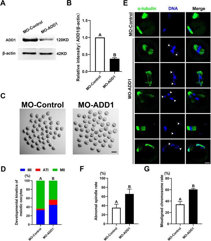
Result: We found that the expression levels and localization of ADD1, S726 phosphorylated ADD1 (p-ADD1), and TPX2 proteins exhibited spindle assembly-dependent dynamic changes during mouse oocyte meiosis. Taxol treatment, which stabilizes the microtubule polymer and protects it from disassembly, made the signals of ADD1, p-ADD1, and TPX2 present in the microtubule organizing centers of small asters and spindles. Knockdown of approximately 60% of ADD1 protein levels destabilized interpolar microtubules in the meiotic spindle, resulting in aberrant chromosome alignment, reduced first polar body extrusion, and increased aneuploidy in metaphase II oocytes, but did not affect K-fiber homeostasis and the expression and localization of TPX2. Strikingly, TPX2 deficiency caused increased protein content of ADD1, but decreased expression and detachment of p-ADD1 from the spindle, thereby arresting mouse oocytes at the metaphase I stage with collapsed spindles.

Conclusion: Phosphorylation of ADD1 at S726 by TPX2 mediates acentriolar spindle assembly and precise chromosome segregation in mouse oocytes.

Keywords: ADD1; Acentriolar spindle assembly; Aneuploidy; Interpolar microtubule stability; Mouse oocyte; TPX2.


Conflict of interest statement

The authors have no relevant financial or non-financial interests to disclose.

Figures

Fig. 1
Fig. 1
Expression and subcellular localization of ADD1 during mouse oocyte meiotic maturation. A The protein levels of ADD1 in oocytes at the GV, GVBD, MI, ATI, and MII stages were detected by immunoblotting. Protein loading was verified by the detection of β-actin. The molecular mass of target proteins is indicated on the right. B ADD1 was quantified for four independent repeats (normalized to β-actin, arbitrary units). Different lowercase letters above columns denote statistical difference at p < 0.05 by one-way ANOVA with the Tukey test. C Spatial distribution of ADD1 and spindle microtubules in oocytes at different stages. D Colocalization of ADD1 and γ-tubulin in MI and MII oocytes. E Localization of ADD1 in MI and MII oocytes exposed to taxol. F Distribution of ADD1 in MI and MII oocytes treated with nocodazole. Red, ADD1; green, α-tubulin; blue, DNA; pink, γ-tubulin; Merge, overlapping of red, green, blue, and pink. Bar, 20 μm
Fig. 2
Fig. 2
Expression and subcellular localization of p-ADD1 during mouse oocyte meiotic maturation. A The protein levels of p-ADD1 in oocytes at various stages were analyzed by western blot. Protein loading was verified by the detection of β-actin. The molecular mass of target proteins is indicated on the right. B p-ADD1 was quantified for three independent repeats (normalized to β-actin, arbitrary units). Bars that do not share the same lowercase letter are significantly different at p < 0.05 by one-way ANOVA with the Tukey test. C Immunofluorescent localization of p-ADD1 and spindle microtubules in meiotic oocytes at various stages. D Distribution of p-ADD1 in MI and MII oocytes treated with taxol. E Localization of p-ADD1 in MI and MII oocytes exposed to nocodazole. Pink or red, p-ADD1; green, α-tubulin; blue, DNA; Merge, overlapping of pink/red, green, and blue. Bar, 20 μm
Fig. 3
Fig. 3
Effects of ADD1 deficiency on oocyte maturation, spindle assembly, and chromosome alignment. A Immunoblot probed with anti-ADD1 antibody demonstrating the depletion efficiency of ADD1-specific morpholino (MO-ADD1) in mouse oocytes. Protein loading was verified by the detection of β-actin. The molecular mass of target proteins is indicated on the right. B The ADD1 protein levels in control-MO- and ADD1-MO-injected oocytes were quantified for three independent repeats (normalized to β-actin, arbitrary units). In each set of experiments, the protein level of ADD1 was normalized to the value of oocytes in the control group. C Representative images of oocytes cultured in vitro for 16 h after control-MO or ADD1-MO treatment. Bar, 100 μm. D Depletion of ADD1 caused the failure of the first polar body extrusion in mouse oocytes. The rate of oocytes at the MI, AT1, and MII stages was quantified 16 h after meiotic resume in oocytes injected with control-MO and ADD1-MO, respectively. E Spindle morphologies and chromosome alignment 16 h after maturation culture in oocytes injected with control-MO or ADD1-MO. Green, α-tubulin; blue, DNA; Merge, overlapping of green and blue. Bar, 20 μm. F The rate of the oocyte with an aberrant spindle was recorded in the control-MO and ADD1-MO-injected oocytes. G The rate of the oocyte with misaligned chromosomes was quantified in the control and ADD1-depleted oocytes. Each column indicates the mean ± SEM of the three independent repeats in D, F, G. Different uppercase letters above columns denote statistical difference at p < 0.01 by student’s t-test in B, whereas bars that do not share the same uppercase letter are significantly different at p < 0.01 by the chi-square test in D, F, G
Fig. 4
Fig. 4
Loss of interpolar microtubules in ADD1-depleted oocytes. A Immunofluorescence images of the MII spindle in calcium-treated control and ADD1-depleted mouse oocytes. Green, α-tubulin; blue, DNA; Merge, overlapping of green and blue. Enlarge, an enlarged view of the image in the white boxes of the merged image. Bar, 20 μm. B Quantification of total fluorescence intensity (arbitrary units) of spindle microtubules in calcium-treated control and ADD1-depleted mouse oocytes at the MII stage. In each set of experiments, the fluorescence intensity of spindle microtubule staining was normalized to the value of oocytes in the control group. Bars that do not share the same uppercase letter are significantly different at p < 0.01 by the student’s t-test
Fig. 5
Fig. 5
Depletion of ADD1 generates aneuploid eggs. A Representative images of euploid and aneuploid eggs from control and ADD1-depleted oocytes, respectively. Chromosomes were stained with DAPI. Scale bar, 5 μm. B The rate of aneuploid eggs was recorded in control and ADD1-depleted oocytes. Bars that do not share the same uppercase letter are significantly different at p < 0.01 by the chi-square test
Fig. 6
Fig. 6
Expression and subcellular localization of TPX2 during mouse oocyte meiotic maturation. A The protein content of TPX2 in oocytes at the GV, GVBD, MI, ATI, and MII stages was detected by immunoblotting. Protein loading was verified by the detection of β-actin. The molecular mass of target proteins is indicated on the right. B TPX2 was quantified for four independent repeats (normalized to β-actin, arbitrary units). Different lowercase letters above bars indicate statistical difference at p < 0.05 by ANOVA with the Tukey test. C Immunofluorescent localization of TPX2 in meiotic oocytes at various stages. D Spatial distribution of TPX2 and spindle microtubules in oocytes at different meiotic maturation stages. E Oocytes at the MI and MII stages were treated with taxol and then double-stained for TPX2 and α-tubulin. F Oocytes at the MI and MII stages were exposed to nocodazole and then co-stained for p-ADD1 and α-tubulin. Red, ADD1; green, α-tubulin; blue, DNA; Merge, overlapping of red, green, and blue. Bar, 20 μm
Fig. 7
Fig. 7
Effects of TPX2 depletion on oocyte maturation, spindle assembly, chromosome alignment, and the expression of p-ADD1 and ADD1. A Immunoblot probed with anti-TPX2 antibody demonstrating the depletion efficiency of TPX2-specific morpholino (MO-TPX2) in mouse oocytes. B TPX2 protein content in control-MO and TPX2-MO-injected oocytes was quantified for three independent repeats. C Representative images of oocytes cultured in vitro for 16 h after treatment with control-MO or TPX2-MO. Bar, 100 μm. D Depletion of TPX2 caused meiotic cell cycle arrest in MI in mouse oocytes. E Subcellular localization of p-ADD1, spindle morphologies, and chromosome alignment after 16 h of maturation culture in oocytes injected with control-MO or TPX2-MO. Bar, 20 μm. F The rate of the oocyte with a normal spindle, aberrant spindle, or no spindle was quantified in the control-MO and ADD1-MO-injected oocytes. G The rate of the oocyte with normal p-ADD1 localization, abnormal p-ADD1 localization, or no p-ADD1 localization was recorded in the control and ADD1 disrupted oocytes. H The depletion of TPX2 downregulated the phosphorylation of ADD1 at S726 in mouse oocytes. I The protein level of p-ADD1 in control and TPX2-depleted oocytes was quantified for three independent repeats. J Effect of TPX2 depletion on ADD1 protein expression in mouse oocytes. (K) ADD1 protein levels in control-MO and TPX2-MO-injected oocytes were quantified for three independent replicates. In B, I, K, each bar denotes the mean ± SEM of the three independent repeats, and different uppercase letters or lowercase letters above columns indicate statistical difference at p < 0.01 or p < 0.05 by student’s t-test, respectively. Whereas bars that do not share the same uppercase letter are significantly different at p < 0.01 by the chi-square test in D, F, G
Fig. 8
Fig. 8
Schematic illustration of the molecular mechanism by which ADD1 regulates spindle assembly in mouse oocytes. ADD1 is phosphorylated by TPX2 and aggregates to the spindle poles to regulate the homeostasis of interpolar microtubules to ensure the formation of functional spindles and the proper segregation of chromosomes in mouse oocytes

Similar articles

Cited by

References

    1. Hassold T, Hunt P. To err (meiotically) is human: the genesis of human aneuploidy. Nat Rev Genet. 2001;2(4):280–291. doi: 10.1038/35066065. - DOI - PubMed
    1. Blengini CS, Schindler K. Acentriolar spindle assembly in mammalian female meiosis and the consequences of its perturbations on human reproduction dagger. Biol Reprod. 2022;106(2):253–263. doi: 10.1093/biolre/ioab210. - DOI - PMC - PubMed
    1. Mogessie B, Scheffler K, Schuh M. Assembly and positioning of the oocyte meiotic spindle. Annu Rev Cell Dev Biol. 2018;34:381–403. doi: 10.1146/annurev-cellbio-100616-060553. - DOI - PubMed
    1. Battaglia DE, Goodwin P, Klein NA, Soules MR. Influence of maternal age on meiotic spindle assembly in oocytes from naturally cycling women. Hum Reprod. 1996;11(10):2217–2222. doi: 10.1093/oxfordjournals.humrep.a019080. - DOI - PubMed
    1. Wang WH, Sun QY. Meiotic spindle, spindle checkpoint and embryonic aneuploidy. Front Biosci. 2006;11:620–636. doi: 10.2741/1822. - DOI - PubMed

LinkOut - more resources