

Masson Tumor of the Central Nervous System: A Case Report and Review of Literature
- PMID: 36540012
- PMCID: PMC9793341
- DOI: 10.12659/AJCR.937597
Masson Tumor of the Central Nervous System: A Case Report and Review of Literature
Abstract
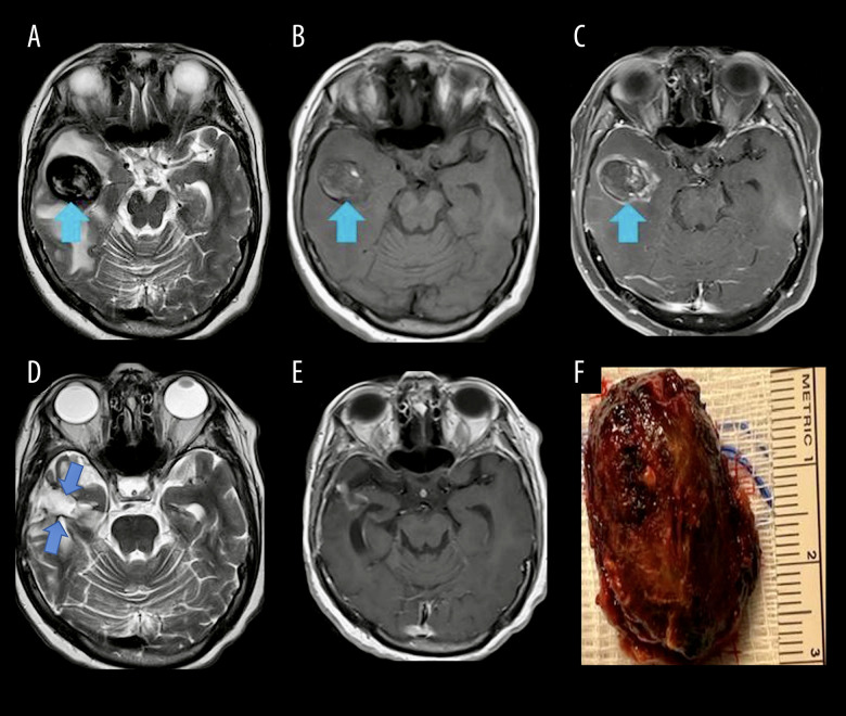
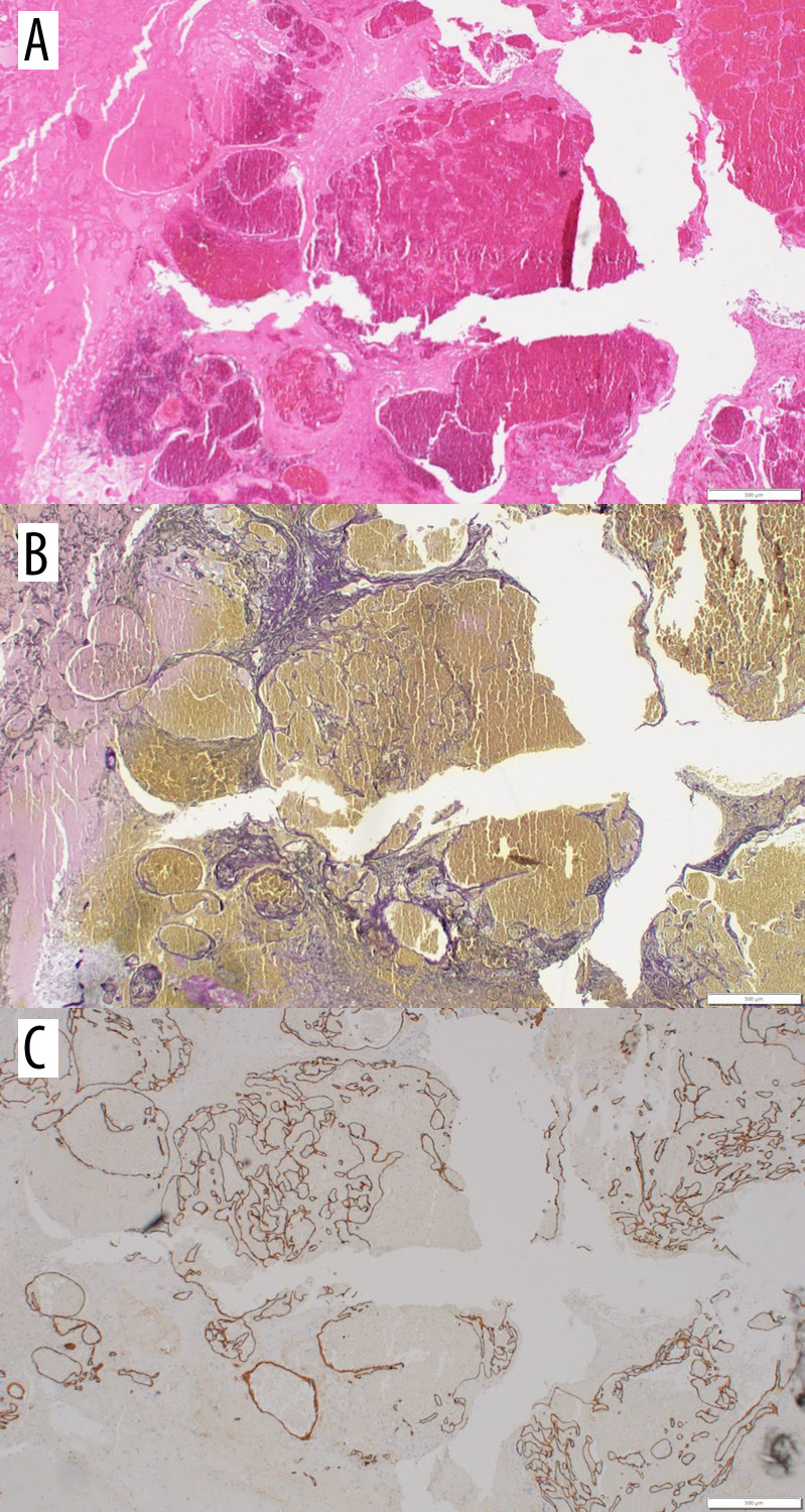
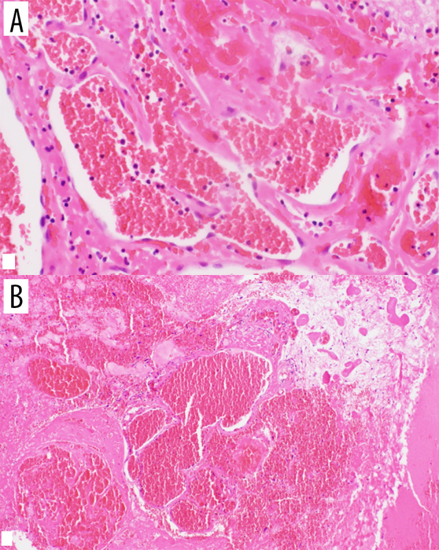
BACKGROUND Masson's tumor, also known as intravascular papillary endothelial hyperplasia (IPEH), is an unusual endothelial proliferation that leads to improper thrombus development due to faulty endothelial structure. Although IPEH is rare in the central nervous system, it can arise at any location in the brain. Headaches, seizures, and focal neurological symptoms ae the most common presenting symptoms. It is more common in females and it can occur at any age. CASE REPORT Herein, we present a 65-year-old female patient with a progressively enlarging right temporal lobe mass that was initially considered metastatic ovarian carcinoma. She underwent a right temporal craniotomy and the lesion was totally resected. Contrary to expectations, the pathology report was an IPEH. CONCLUSIONS In this paper, we conducted a literature review of previously reported cerebral IPEH cases, with a focus on their clinical and radiological presentations, management, and especially their association with previous radiotherapy. The important point is that one-third of the cases had a history of radiation therapy to the head, and most of them had stereotactic radiosurgery (SRS) on the location of the brain from which IPEH subsequently developed. The major question for which we are looking for an answer is its relationship with previous radiotherapies. We wanted to know how many of these cases were associated with radiotherapy in the same area, the time interval from radiotherapy to the onset of IPEH or symptoms, the dose of the previous radiotherapy, and, overall, if there is any cause-effect relationship between IPEH and radiotherapy.
Conflict of interest statement
Figures
References
-
- Cagli S, Oktar N, Dalbasti T, et al. Intravascular papillary endothelial hyper-plasia of the central nervous system – four case reports. Neurol Med Chir (Tokyo) 2004;44(6):302–10. - PubMed
-
- Enzinger F, Enzinger WS. Weiss’s soft tissue tumors. Shock. 2008;30(6):754.
-
- Prat GP, Jimenez MS, Caro PC, et al. Case report and review of the literature. World Neurosurgery. 2018;114:194–203. - PubMed
-
- Karamchandani J, Vogel H, Fischbein N, et al. Extravascular papillary endothelial hyperplasia mimicking neoplasm after radiosurgery: Case report. Neurosurgery. 2012;70(4):E1043–48. - PubMed
-
- Masson P. Hemangioendotheliome vegetant intravasculaire. Bull Soc Anat (Paris) 1923;93:517.
Publication types
MeSH terms
LinkOut - more resources
Full Text Sources
