. 2022 Sep 29;4(24):5257-5269.
doi: 10.1039/d2na00453d. eCollection 2022 Dec 6.

The aspect ratio effect on the cytotoxicity of inert nano-particles flips depending on particle thickness, and is one of the reasons for the literature inconsistency

Affiliations

The aspect ratio effect on the cytotoxicity of inert nano-particles flips depending on particle thickness, and is one of the reasons for the literature inconsistency

Yifat Brill-Karniely et al. Nanoscale Adv. .

Abstract

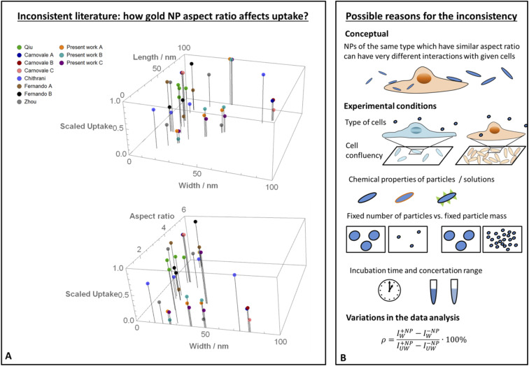
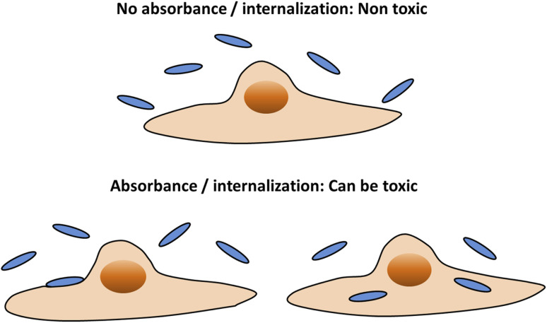
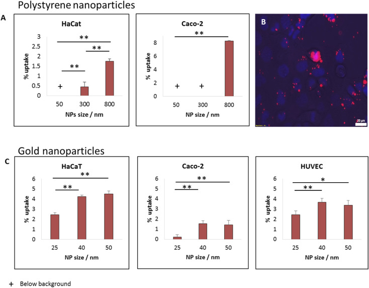
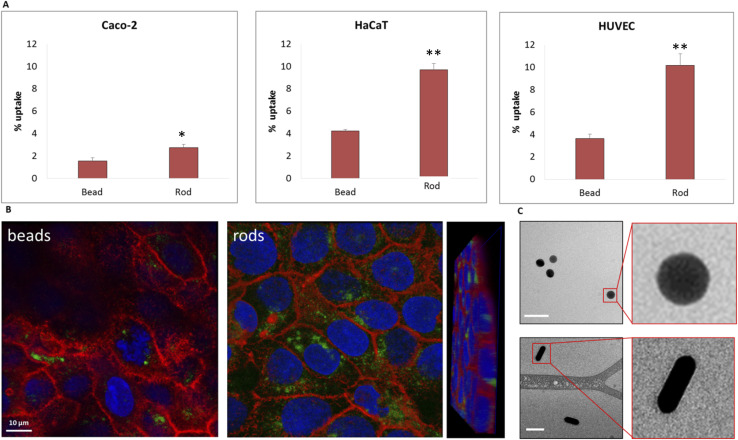
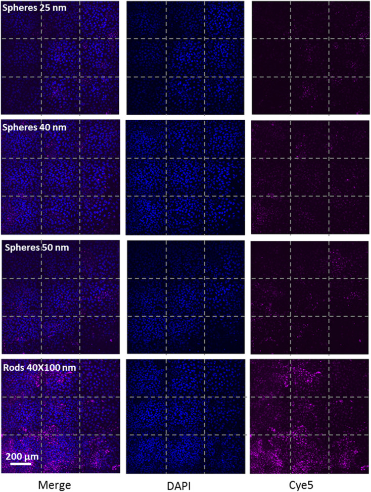
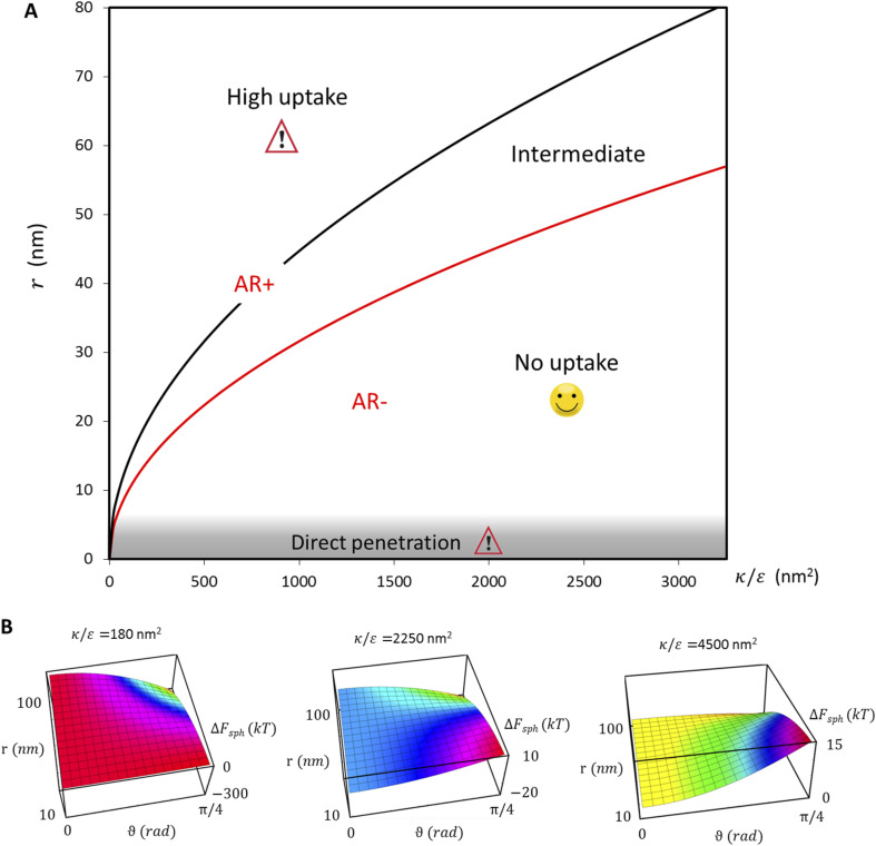
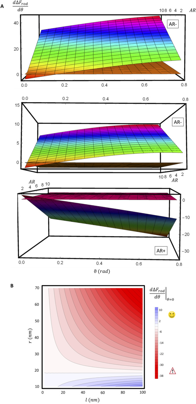
The interaction of inert nano-particles with cells has significant effect on the potential cytotoxicity of the particles. The role of particle aspect ratio in the interaction with cells was largely studied in the literature; however non consistent conclusions were obtained. In the present study a detailed physical model is presented as well as a set of experimental work and a scan of literature data. The aim was to investigate the role of particle size and aspect ratio in cell uptake, and to examine possible sources of the literature inconsistency. Cells which provide the first line of contact with particles in the human body were incubated with seven types of particles. These included spherical and rod gold nanoparticles, as well as larger spherical polystyrene particles in various sizes. We stress that in order to achieve comparative insight careful attention needs to be given to the experimental conditions and to the data analysis. Furthermore, our physical model shows that conclusions regarding the role of aspect ratio in NP uptake largely depend on the radius of the particles. The aspect ratio cannot be regarded as a sole geometrical parameter which determines the interaction of inert nano-particles with cells. When discussing particles larger than 10 nm (for which passive diffusion is irrelevant), the effect of the aspect ratio flips depending on the particle thickness. For particles thicker than ∼35 nm, the longer they are the more toxic they would be, however this trend opposes for thinner NPs, where larger aspect ratio results in reduced uptake and toxicity. Therefore, rod non-functionalized particles whose thickness is between 15 and 30 nm, and are relatively long, are expected to be the safest, with minimal cytotoxicity.


Conflict of interest statement

There are no conflicts to declare.

Figures

Fig. 1
Fig. 1. Literature inconsistency. (A) A comparison between results published in different studies (including the present work), which examined how the uptake of gold NPs by cells was affected by the particles aspect ratio. In each experiment, the highest uptake was scaled to 1 and other data points from the same experiment were normalized accordingly. The scaled data is shown as a function of the width and length of the particles (top), or as a function of the width and aspect ratio (bottom). In both of these presentations, it is clear that the trends were non-consistent. (B) Proposed reasons for the literature inconsistency. A conceptual point is that the aspect ratio cannot be regarded as the sole geometrical parameter; in the present work we show that AR effect flips depending on the NP radius. Other explanations include different behavior of cell types, variations in experimental conditions such as cell confluency, chemical properties of the particles, different definitions of fixed NP amount, variations in incubation time, concentration range and also differences in the data analysis.
Fig. 2
Fig. 2. To maximize the potential safety of nanoparticles, it would be desirable that the particles would not cause either cell death or reduction in functionality. A main step towards this goal would be to prevent physical interaction between the particles and the cells. For that aim both NP adsorption to the cell membrane, and NP internalization into the cells would be non-desirable.
Fig. 3
Fig. 3. Effect of particle size on the uptake of beads, for a wide range of nano–sub micron particle size. For either polystyrene or gold particles, smaller NPs (above the passive diffusion scale) were less uptaken by cells and are thus expected to be less toxic. (A) Percentage of uptake of polystyrene beads in HaCat and CaCo2 cells. (B) Snapshot of CaCo2 cells after 24 h incubation with 800 nm beads. (C) Gold NPs uptake in HaCat, CaCo2 and HUVEC cells. All uptake measurements were done using a Microplate Reader after 24 hours of incubation with the particles. NP concentrations: 25 or 12.5 μg ml−1 for the polystyrene or gold particles, respectively. p-value: ** ≤0.001, * ≤0.01.
Fig. 4
Fig. 4. The uptake of 40 nm gold beads is lower than that of 40 × 100 nm rods, thus the beads are expected to be less toxic. The gold NPs were incubated with CaCo2, HaCat and HUVEC cells for 24 hours. (A) Uptake results from Microplate Reader measurements and (B) Confocal images of HaCat cells after 24 h incubation with the particles. NP concentration was 12.5 μg ml−1. (C) Cryo TEM images confirmed that the particles did not aggregate. p-value: ** ≤0.001, * ≤0.01.
Fig. 5
Fig. 5. Fields of spinning disc confocal images confirmed that the uptake of spherical gold NPs increased with NP size from 25 to 40 nm diameter, and that the uptake of 40 × 100 nano-rods was further dramatically higher. The particles were incubated with HaCat cells for 24 h.
Fig. 6
Fig. 6. Illustration of physical model geometry in the case of (A) parallel or (B) tilted contact. Red and yellow lines represent the contact areas between the membrane and the particle within the cylindrical regime or the cap margins of the NP, respectively. The black line represents bent membrane not in contact with the NP. Definitions of the variables and parameters are described in the main text.
Fig. 7
Fig. 7. (A) Phase diagram summarizing uptake patterns of non-functionalized particles, and the consequence effects on cytotoxicity, as derived from our physical model and elaborated in detail in the main text. Importantly, we found that the effect of aspect ratio depends on the radius of the NPs. The red curve shows the separation between the regimes AR+ and AR−, at which elevated aspect ratio increases or decreases the uptake, respectively. Moreover, we found a finer separation into three phases in the investigation of sphere uptake as a reference: (i) when , ΔFsph(θ) is an increasing function thus particles are expected not to physically interact with cells and would therefore be safe for use. (ii) In the intermediate regime the interaction of the NPS with the cells would not be spontaneous, with a barrier in the free energy, thus uptake may occur depending on factors such as the cell type, the adhesion strength and temperature. (iii) For the free energy would be a decreasing function thus large interaction between the NPs and the cells is expected, potentially leading to large cytotoxicity. In addition, for NPs whose radius is smaller than 5 nm passive diffusion mechanisms would be relevant. In this case, which was beyond the scope of the present model, large NP uptake is expected, leading to cytotoxicity. (B) Surface plots showing ΔFsph for three values of demonstrate that smaller particles (above the direct passive diffusion) are expected to be the safest (with minimal uptake); and that the safety of particles further depends on , whereas the lower is this ratio the “easier” it is for cells to uptake particles.
Fig. 8
Fig. 8. (A) Derivative of ΔFrod with respect to θ, as a function of θ and of the aspect ratio AR. Typical values of 0.002kBT/nm2 and 2.3kBT, respectively, were used for ε and κ. The plots correspond to three values of r: 20 nm (top), and 40 nm (middle) – which are smaller than r*, and 60 nm (bottom) which is larger than r*; a flip from AR− to AR+ is observed between these two regimes. For a clearer view the zero plane was added to the surface plots as a reference. (B) is shown as a function of r and of l. Blue color represents positive values, which indicate on vanishing uptake and higher safety of the rods, and the red color represent negative values and increased probability for cytotoxicity. A threshold in r* ∼ 18 nm, at which the system switches from AR− to AR+ is observed.

Similar articles

Cited by

References

    1. Qiu Y. et al., Surface chemistry and aspect ratio mediated cellular uptake of Au nanorods. Biomaterials. 2010;31(30):7606–7619. doi: 10.1016/j.biomaterials.2010.06.051. - DOI - PubMed
    1. Carnovale C. Bryant G. Shukla R. Bansal V. Identifying Trends in Gold Nanoparticle Toxicity and Uptake: Size, Shape, Capping Ligand, and Biological Corona. ACS Omega. 2019;4:242–256. doi: 10.1021/acsomega.8b03227. - DOI
    1. Chithrani B. Ghazani A. A. Chan W. C. W. Determining the Size and Shape Dependence of Gold Nanoparticle Uptake into Mammalian Cells. Nano Lett. 2006;6(4):662–668. doi: 10.1021/nl052396o. - DOI - PubMed
    1. Fernando D. Sulthana S. Vasquez Y. Cellular Uptake and Cytotoxicity of Varying Aspect Ratios of Gold Nanorods in HeLa Cells. ACS Appl. Bio Mater. 2020;3(3):1374–1384. doi: 10.1021/acsabm.9b00986. - DOI - PubMed
    1. Zhou H. et al., Gold nanoparticles impair autophagy flux through shape-dependent endocytosis and lysosomal dysfunction. J. Mater. Chem. B. 2018;6(48):8127–8136. doi: 10.1039/C8TB02390E. - DOI - PubMed