Case Reports
. 2022 Nov 18;14(11):e31639.
doi: 10.7759/cureus.31639. eCollection 2022 Nov.

Rapidly Progressive Bilateral Vitreoretinal Lymphoma

Affiliations
Case Reports

Rapidly Progressive Bilateral Vitreoretinal Lymphoma

Joseph W Fong et al. Cureus. .

Abstract

A 56-year-old male who presented with unilateral localized sub-retinal lesions suspicious for primary vitreoretinal lymphoma (PVRL) developed florid bilateral ocular involvement and was found to have lesions on MRI of the brain in a five-week period despite the absence of vitreous involvement during the entire course of his disease. His ocular lesions were monitored while on systemic treatment and an excellent clinical response was achieved. His central nervous system (CNS) lesions, however, continued to progress despite chemotherapy and whole-brain radiation. He died 12 months from his time of ocular diagnosis. To our knowledge, this case represents the most rapid progression of PVRL reported in the literature - from unilateral, localized lesions in the sub-retinal space to bilateral ocular involvement and identification of CNS involvement in a five-week period. This case highlights the potential for rapid ocular progression of PVRL stressing the need for early diagnosis. Therefore, we recommend prompt vitreous and, if necessary, sub-retinal biopsy in cases of suspected vitreoretinal lymphoma in addition to neuro-imaging. We emphasize the importance of coordination between pathologists, ophthalmologists, and oncologists for prompt, accurate diagnosis. Delay in diagnosis and treatment can result in rapid intraocular progression and central nervous system spread.

Keywords: central nervous system lymphoma; ocular lymphoma; primary vitreoretinal lymphoma; rapid progression lymphoma; vitreoretinal surgery.


Conflict of interest statement

The authors have declared that no competing interests exist.

Figures
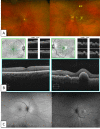
Figure 1
Figure 1. Initial Presentation
On initial presentation, the left fundus reveals multiple creamy yellow retinal lesions (A) that were localized to the sub-RPE space by OCT (B). Autofluorescence (C) and OCT demonstrate a lack of involvement of the right eye. OCT: Optical coherence tomography
Figure 2
Figure 2. Five Weeks Later
(A) Repeat exam of the right fundus reveals no visible retinal lesions one month after the initial presentation, but the left fundus shows a rapid progression of lesions. Repeat OCT (B) and autofluorescence (C) demonstrated new sub-RPE lesions in the previously unaffected right eye and the progression of lesions in the left eye. OCT: Optical coherence tomography
Figure 3
Figure 3. Cytospin Preparation and Flow Cytometry Immunophenotyping of Sub-Retinal Biopsy Specimen
(A) Cytospin preparation of the sub-retinal lesion. There are large atypical lymphoid cells with oval to convoluted nucleus and moderate amount of basophilic cytoplasm. These cells are associated with cell debris and necrosis, as seen in the lower aspect of the image (Wright-Giemsa stain, 60X). (B) Flow cytometry immunophenotyping of the sub-retinal lesion. Top: The large lymphoma cells have intermediate to high scatter and are bright for CD45 (blue population). The large population in gray consisted of cellular debris and had non-specific staining with the other antibodies (not shown). The lymphoma cells are positive for CD19 and CD20, confirming their B-cell lineage. Middle panel: These cells are negative for CD5 and for CD10. Bottom panel: The lymphoma cells demonstrate kappa light chain restriction.
Figure 4
Figure 4. MRI of the Brain with Evidence of Intracranial Disease
T1-weighted MRI of the brain with contrast showing a lenticular lesion within the left anterior frontal lobe (A, left) and a 2.3 x 1.6 cm lesion within the left cerebellar peduncle (B, right).
Figure 5
Figure 5. Serial Exams During Treatment Course
Repeat imaging at one month (A) and three months (B) of systemic chemotherapy following treatment demonstrating excellent response.

References

    1. Primary intraocular diffuse large B-cell lymphoma: diagnostic difficulties in deep retinal infiltrations with vitritis. Kapelko-Slowik K, Urbaniak-Kujda D, Turno-Krecicka A, Potoczek S, Dybko J, Biernat M, Slowik M. Indian J Hematol Blood Transfus. 2016;32:143–147. - PMC - PubMed
    1. Primary intraocular lymphoma: diagnosis and differential diagnosis. Sen HN, Bodaghi B, Hoang PL, Nussenblatt R. Ocul Immunol Inflamm. 2009;17:133–141. - PMC - PubMed
    1. Clinical features, laboratory investigations, and survival in ocular reticulum cell sarcoma. Freeman LN, Schachat AP, Knox DL, Michels RG, Green WR. Ophthalmology. 1987;94:1631–1639. - PubMed
    1. Primary intraocular lymphoma (ocular reticulum cell sarcoma) diagnosis and management. Char DH, Ljung BM, Miller T, Phillips T. Ophthalmology. 1988;95:625–630. - PubMed
    1. Ten-year experience with primary ocular 'reticulum cell sarcoma' (large cell non-Hodgkin's lymphoma) Siegel MJ, Dalton J, Friedman AH, Strauchen J, Watson C. Br J Ophthalmol. 1989;73:342–346. - PMC - PubMed

Publication types

LinkOut - more resources