

Treatment of Merkel Cell Carcinoma Using Intraoperative Flow Cytometry (iFC) Protocol, Optimized for Head and Neck Lesions, as a Means for Tumor Margin Evaluation - a Case Presentation
- PMID: 36540589
- PMCID: PMC9720646
- DOI: 10.26574/maedica.2022.17.3.726
Treatment of Merkel Cell Carcinoma Using Intraoperative Flow Cytometry (iFC) Protocol, Optimized for Head and Neck Lesions, as a Means for Tumor Margin Evaluation - a Case Presentation
Abstract
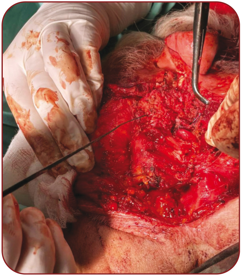
Background: We present the case of a patient with a Merkel cell carcinoma (MCC) of the left preauricular area. Case presentation:A 84-year-old Greek man was examined at the outpatient ENT Department of our clinic with a lesion in the preauricular area that had appeared four months ago. The patient history included antihypertensive and antihyperlipidemic therapy as well as treatment for dementia. The excision of the skin lesion was performed under local anesthesia. The histological examination revealed a Merkel cell carcinoma. The patient underwent a computed tomography (CT) scan that showed a lesion with clear limits in the left parotid gland and lymph nodes. Under general anesthesia, he underwent a left superficial parotidectomy, left submandibular gland excision and radical neck dissection. Histological preparations were analyzed using an intraoperative flow cytometry (iFC) protocol. A radiation therapy concluded the patient's treatment. Conclusion:Even if MCC appears as a less common and more aggressive skin cancer type, a clinician always has to include it in the differential diagnosis of a skin lesion. We found the use of iFC very useful for the diagnosis of this skin cancer.
Figures


References
-
- Swann MH, Yoon J. Merkel Cell Carcinoma. S. eminars in Oncology. 2007;34:51–56. - PubMed
-
- Boyle F, Pendelebury S, Bell D. Further insights into the natural history and management of primary cutaneous neuroendocrine (Mercel Cell carcinoma) carcinoma. Int J Radiat Oncol Biol Phys. 1995;31:315–323. - PubMed
-
- Schwartz RA, Lambert WC. The Merkel cell carcinoma: a 50-year retrospect. J Surg Oncol. 2005;89:5. - PubMed
-
- Medina-Franco H, Urist MM, Fiveash J, et al. Multimodality Treatment of Merkel Cell Carcinoma: Case Series and Literature Review of 1024 Cases. Ann Surg Oncol. 2001;8:204–208. - PubMed
Publication types
LinkOut - more resources
Full Text Sources
Research Materials
Miscellaneous