

WRN helicase and mismatch repair complexes independently and synergistically disrupt cruciform DNA structures
- PMID: 36541070
- PMCID: PMC9890227
- DOI: 10.15252/embj.2022111998
WRN helicase and mismatch repair complexes independently and synergistically disrupt cruciform DNA structures
Abstract
The Werner Syndrome helicase, WRN, is a promising therapeutic target in cancers with microsatellite instability (MSI). Long-term MSI leads to the expansion of TA nucleotide repeats proposed to form cruciform DNA structures, which in turn cause DNA breaks and cell lethality upon WRN downregulation. Here we employed biochemical assays to show that WRN helicase can efficiently and directly unfold cruciform structures, thereby preventing their cleavage by the SLX1-SLX4 structure-specific endonuclease. TA repeats are particularly prone to form cruciform structures, explaining why these DNA sequences are preferentially broken in MSI cells upon WRN downregulation. We further demonstrate that the activity of the DNA mismatch repair (MMR) complexes MutSα (MSH2-MSH6), MutSβ (MSH2-MSH3), and MutLα (MLH1-PMS2) similarly decreases the level of DNA cruciforms, although the mechanism is different from that employed by WRN. When combined, WRN and MutLα exhibited higher than additive effects in in vitro cruciform processing, suggesting that WRN and the MMR proteins may cooperate. Our data explain how WRN and MMR defects cause genome instability in MSI cells with expanded TA repeats, and provide a mechanistic basis for their recently discovered synthetic-lethal interaction with promising applications in precision cancer therapy.
Keywords: Werner; biochemistry; cancer; mismatch repair; synthetic lethality.
© 2022 The Authors. Published under the terms of the CC BY NC ND 4.0 license.
Figures
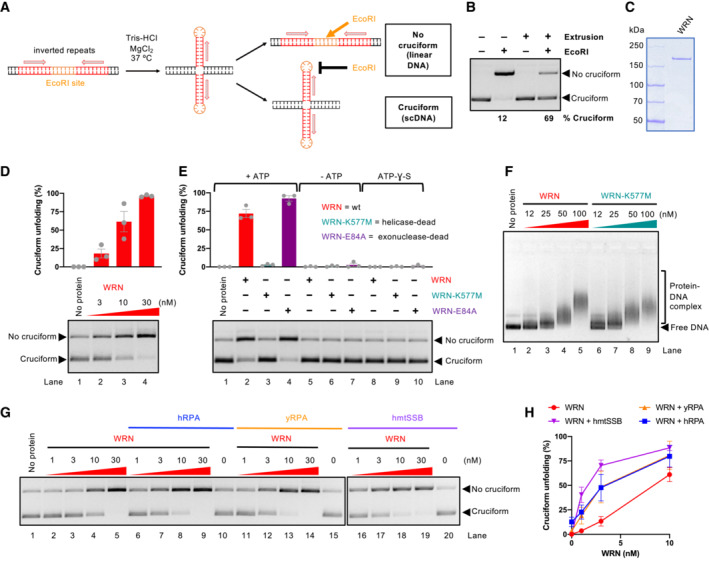
A schematic representation of the cruciform detection assay.
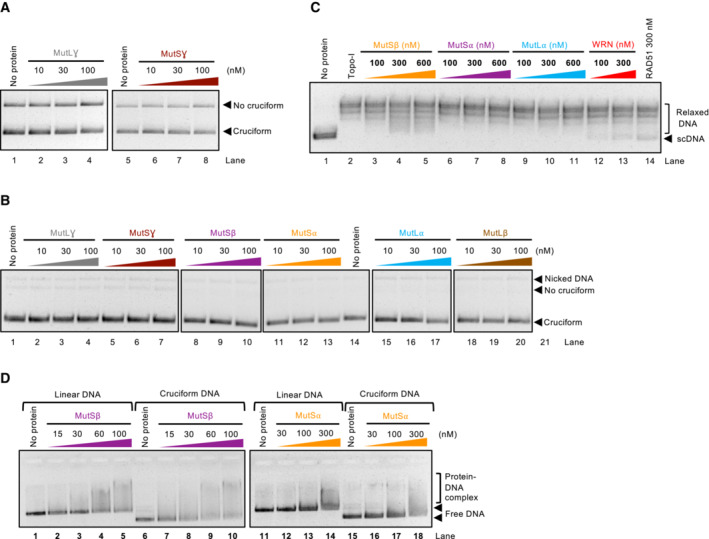
Representative cruciform detection assay. Supercoiled DNA (cruciform) and linear DNA (no cruciform) were resolved on a 1% agarose gel, stained with GelRed. The % of cruciform‐containing molecules are expressed as averages; n = 3.
Representative polyacrylamide gel showing purification of recombinant WRN. The gel was stained with Coomassie Brilliant blue.
Cruciform unfolding assay with increasing WRN concentrations and cruciform consisting of random inverted repeats (random‐IR). Top, quantitation of cruciform unfolding. The amount of linear DNA from the “No protein” lane was subtracted from all other samples. Averages shown; n = 3 technical replicates; error bars, SEM.
Cruciform unfolding assays as in (D) with WRN, WRN‐K577M, and WRN‐E84A (all used at 10 nM), carried out either in the presence or absence of ATP, or with ATP‐ƔS. Averages shown; n ≥ 3 technical replicates; error bars, SEM.
Electrophoretic mobility shift assays with WRN and WRN‐K577M, using pUC19 with the random‐IR cruciform structure as a substrate.
Representative cruciform unfolding assays with increasing concentrations of WRN, together with human RPA (hRPA, 30 nM), yeast S. cerevisiae RPA (yRPA, 30 nM), or human mitochondrial SSB (hmtSSB, 50 nM). pUC19 with the random‐IR cruciform structure was used as a substrate.
Quantitation of assays such as in (G). The amount of linear DNA from the “No protein” lane was subtracted from all other samples. Averages shown; n ≥ 3 technical replicates; error bars, SEM.
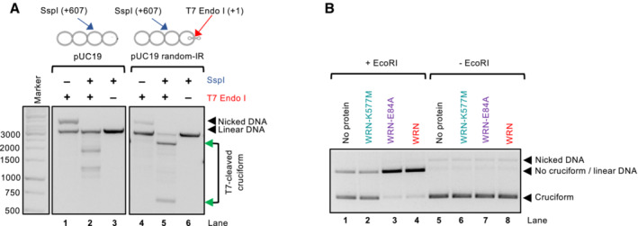
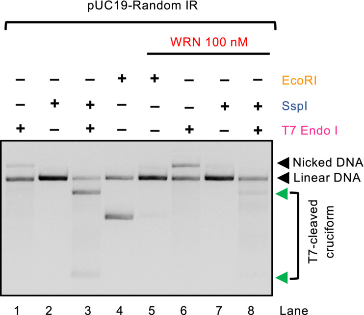
Representative assay showing the activity of T7 Endonuclease I, which cleaves the 4‐way junction formed at the cruciform DNA site. Incubation with T7 Endonuclease I and SspI results in the production of bands at the expected positions of 607 and 2,119 bp, indicated by the green arrows. The SspI restriction site is 607 bp away from the cruciform site. A plasmid without the inverted repeats is cleaved less efficiently and produces bands of different sizes (lane 2), resulting from T7 Endonuclease I activity at other sites that spontaneously extrude in pUC19 DNA. The DNA molecules were resolved by standard electrophoresis on a 1% native agarose gel, stained with GelRed.
Representative cruciform unfolding assay with WRN, WRN‐K577M, and WRN‐E84A (all used at 10 nM). The assay was incubated either with or without EcoRI and the DNA species were separated on a 1% native agarose gel, stained with GelRed.
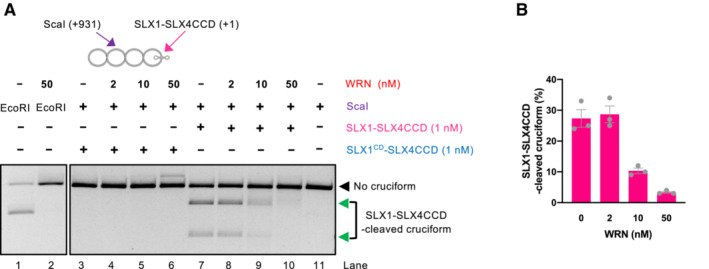
Representative assays with cruciform DNA (consisting of random‐IR) either without or with WRN incubation prior to cruciform detection by SLX1‐SLX4CCD. The ScaI restriction site is 931 bp away from the cruciform site cleaved by SLX1‐SLX4CCD. Combined activity of ScaI and SMX results in 931 and 1,795 bp bands indicated by the green arrows. The intensity of these bands decreases upon WRN incubation, resulting predominantly in linear DNA cut by ScaI, suggesting that WRN removes the substrate for SLX1‐SLX4CCD. The catalytic dead version of SLX1‐SLX4CCD (SLX1CD‐SLX4CCD), where SLX1 carries an R41A mutations in its nuclease domain, has no activity on the cruciform DNA.
Quantitation of assays such as in (A). Averages shown; n = 3 technical replicates; error bars, SEM.
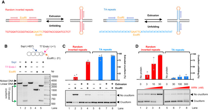
Comparison of cruciform substrates based on random inverted repeats (left) and TA repeats (right). Top, a schematic cartoon; Bottom, DNA sequence.
Mapping of TA repeats cruciform structure. T7 Endonuclease I activity, followed by SspI, cleaves the cruciform within negatively supercoiled DNA, producing two bands of the expected size (607 and 2,119 bp, indicated by the green arrows, lane 2). Simultaneous incubation of the substrate with SspI and EcoRI allows EcoRI cutting at the TA repeats sequence (lane 5), while the extruded cruciform is refractory to EcoRI digestion (lane 4, the * indicates scDNA).
Representative cruciform detection assays. Plasmid DNA was subjected to Cruciform extrusion procedure, where indicated. Supercoiled DNA (cruciform, refractory to EcoRI) and linear DNA (no cruciform, cleaved by EcoRI) were resolved on a 1% agarose gel, stained with GelRed. Top, quantitation of extruded cruciform in % with respect to the amount of total DNA that is present in the lane. Averages shown; n = 4 technical replicates; error bars, SEM.
Representative cruciform unfolding assays with increasing WRN concentrations, using pUC19 with either random inverted repeats or TA repeats as a substrate. Top, quantitation of cruciform unfolding. The amount of linear DNA from the “No protein” lane was subtracted from all other samples. Averages shown; n = 3 technical replicates; error bars, SEM.
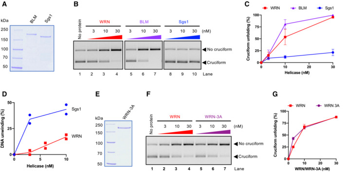
Recombinant BLM and S. cerevisiae Sgs1 used in this study. The gel was stained with Coomassie Brilliant blue.
Representative cruciform unfolding assay with increasing concentrations of WRN, BLM, and Sgs1. Cruciform DNA consisting of random inverted repeats was used.
Quantitation of assays such as (B). Averages shown; n ≥ 3 technical replicates; error bars, SEM.
Quantitation of DNA unwinding from experiments such shown in (EV4D). Averages and individual data points shown; n = 2 technical replicates.
Recombinant WRN‐3A (S991A T1152A S1256A) used in this study. The gel was stained with Coomassie Brilliant blue.
Representative cruciform unfolding assays with increasing concentrations of WRN and WRN‐3A. Cruciform DNA consisting of random inverted repeats was used.
Quantitation of assays such as in (F). Averages shown; n = 3 technical replicates; error bars, SEM.
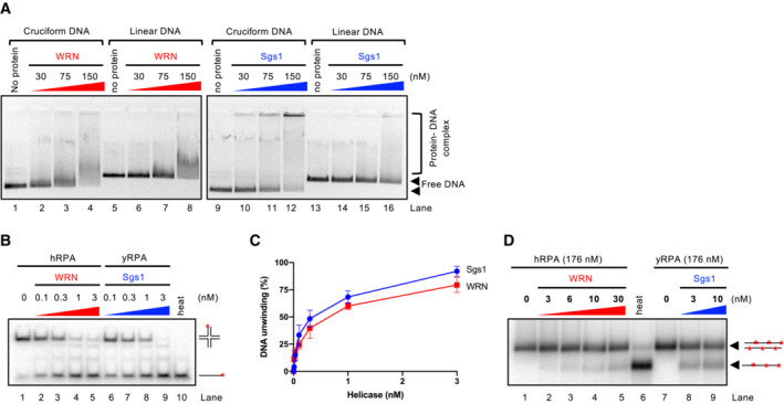
Representative electrophoretic mobility shift assay with WRN and Sgs1, using either pUC19 harboring the random inverted repeat‐based cruciform structure (circular), or linearized pUC19. The samples were run on a 0.8% unstained native agarose gel. The gel was stained after the run with GelRed.
Representative helicase assay with increasing concentrations of WRN and Sgs1, using an oligonucleotide‐based Holliday junction as a substrate. Reactions were supplemented with either human or yeast RPA (15 nM) and analyzed by 10% native acrylamide gel electrophoresis. Red asterisk indicates the position of the radioactive label.
Quantitation of DNA unwinding from assays such as in (B). Averages shown; n ≥ 3; error bars, SEM.
Representative helicase assay with increasing concentrations of WRN and Sgs1, using a 2.2 kbp‐long dsDNA substrate. Reactions were supplemented with either human or yeast RPA and analyzed by electrophoresis on a 1% agarose gel.

Primary structure of WRN. Full‐length and truncation WRN variants are shown.
Recombinant full‐length and truncation WRN variants used in this study are shown. The gel was stained with Coomassie Brilliant blue.
Quantitation of DNA unwinding from assays such as shown in (EV5). Averages shown; n = 3 technical replicates; error bars, SEM.
Cruciform unfolding assays with increasing concentrations of WRN variants.
Quantitation of assays such as shown in (D). Averages shown; n = 3 technical replicates; error bars, SEM.

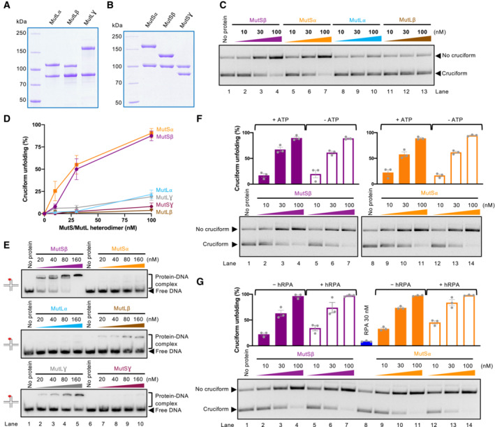
MutL homolog heterodimers used in this study: MutLα (MLH1‐PMS2), MutLβ (MLH1‐PMS1), and MutLγ (MLH1‐MLH3).
MutS homolog heterodimers used in this study: MutSα (MSH2‐MSH6), MutSβ (MSH2‐MSH3), and MutSγ (MSH4‐MSH5).
Cruciform unfolding assays with MutSβ, MutS⍺, MutL⍺, and MutLβ. Cruciform DNA based on random inverted repeats was used.
Quantitation of assays such as in (C) and (EV6A). Averages shown; n = 3 technical replicates; error bars, SEM.
Representative electrophoretic mobility shift assays with the MutS and MutL homolog heterodimers, using an oligonucleotide‐based Holliday junction as a substrate. Red asterisk indicates the position of the radioactive label.
Representative cruciform unfolding assays with MutSβ and MutS⍺, carried out without or with ATP, using the random‐IR cruciform as a substrate. Top, quantitation of cruciform unfolding. Averages shown; n = 3 technical replicates; error bars, SEM.
Representative cruciform unfolding assays with MutSβ and MutS⍺, carried out without or with hRPA, using the random‐IR cruciform as a substrate. Top, quantitation of cruciform unfolding. Averages shown; n = 3 technical replicates; error bars, SEM.
Representative cruciform unfolding assay with MutLγ and MutSγ, using the random‐IR cruciform as a substrate.
Representative cruciform unfolding assay with the MutL and MutS homolog heterodimers, carried out in the absence of EcoRI. The reactions indicate that the proteins do not cut DNA under the assay conditions.
Representative Topoisomerase‐I‐coupled supercoiling assay with MutSβ, MutS⍺, MutL⍺, WRN, and RAD51. The reactions were analyzed by standard electrophoresis on a 1% native agarose gel. The gel was stained with GelRed after electrophoresis.
Representative electrophoretic mobility shift assay with MutSβ and MutSα, using either circular pUC19 with the random inverted repeat cruciform structure, or linear pUC19 as a substrate. 0.8% unstained native agarose gel was used to separate the protein and DNA species.
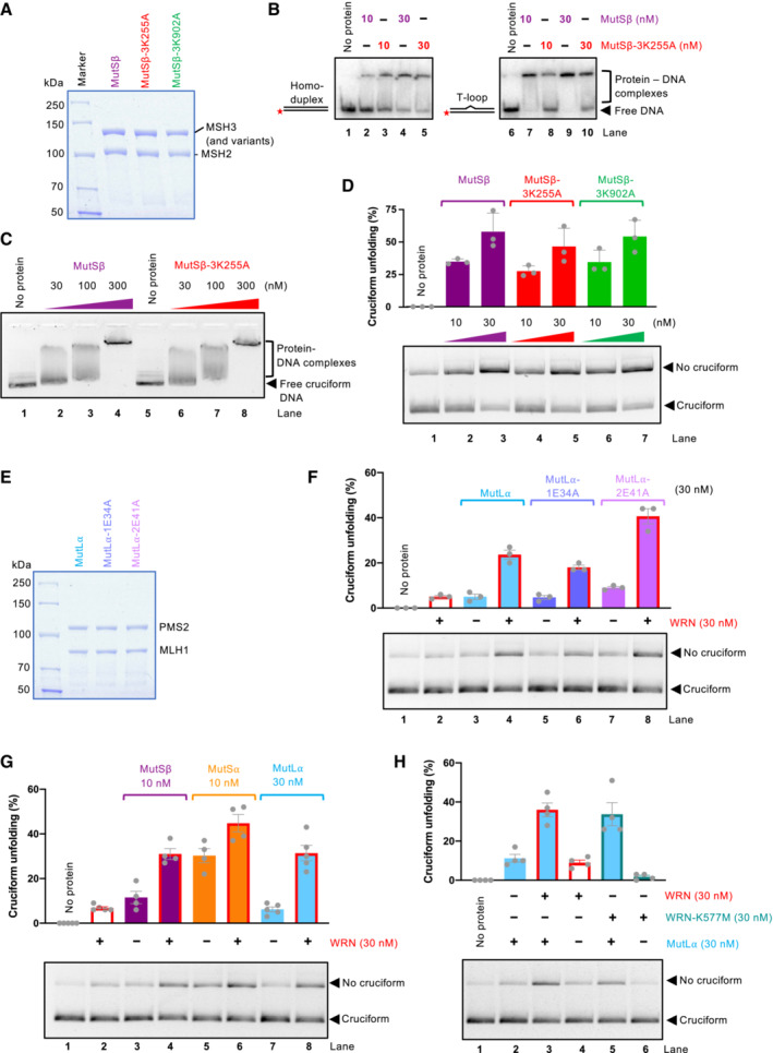
Representative polyacrylamide gel showing recombinant MutSβ, MutSβ‐3K255A (MSH3 with mutation K255A, deficient in mismatch recognition), and MutSβ‐3K902A (MSH3 with mutation K902A, ATPase‐deficient). The gel was stained with Coomassie Brilliant blue.
Electrophoretic mobility shift assay with MutSβ, MutSβ‐3K255A, and MutSβ‐3K902A, using either dsDNA (50 bp) or dsDNA bearing one extrahelical T, as a substrate. 6% native acrylamide gel was used.
Electrophoretic mobility shift assays with MutSβ and MutSβ‐3K255A, using circular pUC19 with the AT repeat cruciform structure, as a substrate. 0.8% native agarose gel was used.
Cruciform unfolding assays with MutSβ, MutSβ‐3K255A, and MutSβ‐3K902A. Bottom, representative experiments; top, quantitation, averages shown; n = 3 technical replicates; error bars, SEM.
Representative polyacrylamide gel showing recombinant MutLα, MutL⍺‐1E34A (MLH1 with mutation E34A, ATPase‐deficient), and MutL⍺‐2E41A (PMS2 with mutation E41A, ATPase‐deficient). The gel was stained with Coomassie Brilliant blue.
Unfolding of TA cruciform by WRN, MutLα, and its ATPase‐deficient variants. Bottom, representative experiments; top, quantitation; averages shown; n = 3 technical replicates; error bars, SEM.
Unfolding of TA cruciform by WRN and MMR proteins. Bottom, representative experiments; top, quantitation; averages shown; n = 4 technical replicates; error bars, SEM.
Unfolding of TA cruciform by MutLα and wild‐type WRN or helicase dead WRN‐K577M. Bottom, representative experiments; top, quantitation, averages shown; n = 4 technical replicates; error bars, SEM.
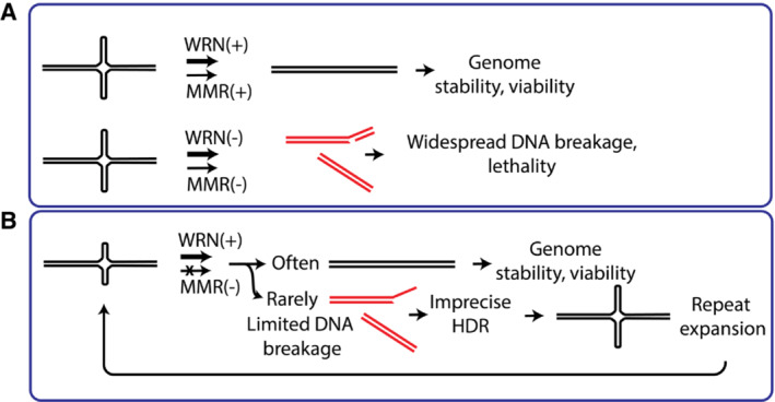
Top: in wild‐type cells (MMR‐ and WRN‐proficient), cruciform DNA is efficiently unfolded, and does not cause any problems. WRN and MMR complex may act together or separately on cruciform DNA. Bottom: in MMR‐deficient (MSI) cells, inactivation of WRN triggers very frequent DNA breaks at cruciform sites, leading to cellular lethality.
In MMR‐deficient (MSI+) but WRN proficient cells, large parts of DNA are generally stable. Only very few cruciform structures are formed in the absence of MMR and subject to breakage. Imprecise homology‐directed repair may lead to repeat expansion. DNA breaks are rare and do not result in lethality. Over many generations, cruciform structures extend at random genomic locations.
References
-
- Ahn B, Harrigan JA, Indig FE, Wilson DM 3rd, Bohr VA (2004) Regulation of WRN helicase activity in human base excision repair. J Biol Chem 279: 53465–53474 - PubMed
-
- Anand R, Ranjha L, Cannavo E, Cejka P (2016) Phosphorylated CtIP functions as a Co‐factor of the MRE11‐RAD50‐NBS1 endonuclease in DNA end resection. Mol Cell 64: 940–950 - PubMed
-
- Anand R, Pinto C, Cejka P (2018) Methods to study DNA end resection I: recombinant protein purification. Methods Enzymol 600: 25–66 - PubMed
Publication types
MeSH terms
Substances
Grants and funding
LinkOut - more resources
Full Text Sources
Other Literature Sources
Miscellaneous
