. 2023 Jan 26;205(1):e0042422.
doi: 10.1128/jb.00424-22. Epub 2022 Dec 21.

LdtC Is a Key l,d-Transpeptidase for Peptidoglycan Assembly in Mycobacterium smegmatis

Affiliations

LdtC Is a Key l,d-Transpeptidase for Peptidoglycan Assembly in Mycobacterium smegmatis

Zanah K Francis et al. J Bacteriol. .

Abstract

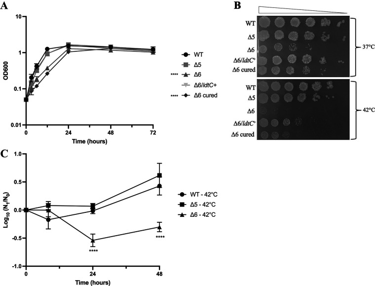
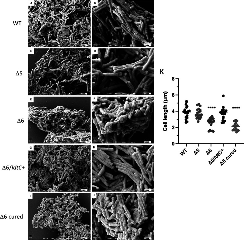
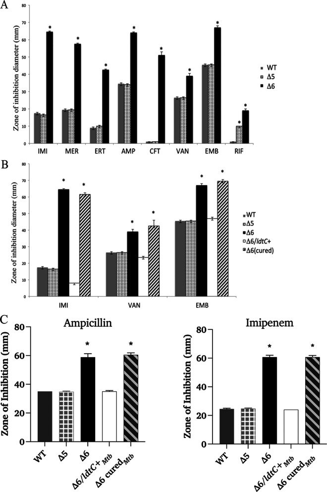
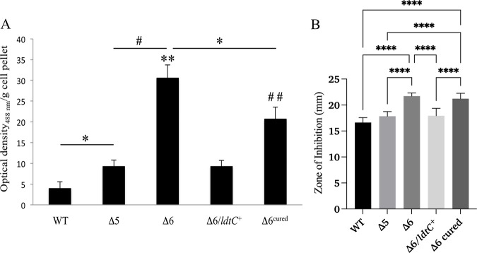
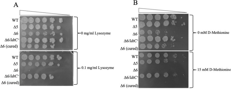
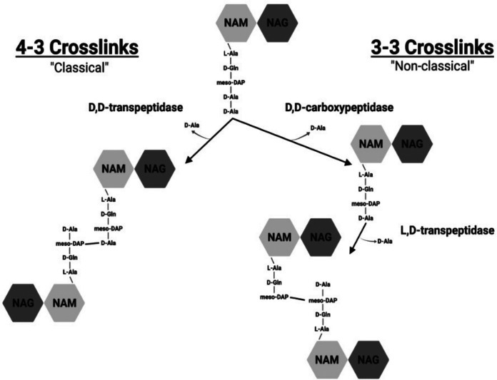
The peptidoglycan of mycobacteria has two types of direct cross-links, classical 4-3 cross-links that occur between diaminopimelate (DAP) and alanine residues, and nonclassical 3-3 cross-links that occur between DAP residues on adjacent peptides. The 3-3 cross-links are synthesized by the concerted action of d,d-carboxypeptidases and l,d-transpeptidases (Ldts). Mycobacterial genomes encode several Ldt proteins that can be classified into six classes based upon sequence identity. As a group, the Ldt enzymes are resistant to most β-lactam antibiotics but are susceptible to carbapenem antibiotics, with the exception of LdtC, a class 5 enzyme. In previous work, we showed that loss of LdtC has the greatest effect on the carbapenem susceptibility phenotype of Mycobacterium smegmatis (also known as Mycolicibacterium smegmatis) compared to other ldt deletion mutants. In this work, we show that a M. smegmatis mutant lacking the five ldt genes other than ldtC has a wild-type phenotype with the exception of increased susceptibility to rifampin. In contrast, a mutant lacking all six ldt genes has pleiotropic cell envelope defects, is temperature sensitive, and has increased susceptibility to a variety of antibiotics. These results indicate that LdtC is capable of functioning as the sole l,d-transpeptidase in M. smegmatis and suggest that it may represent a carbapenem-resistant pathway for peptidoglycan biosynthesis. IMPORTANCE Mycobacteria have several enzymes to catalyze nonclassical 3-3 linkages in the cell wall peptidoglycan. Understanding the biology of these cross-links is important for the development of antibiotic therapies to target peptidoglycan biosynthesis. Our work provides evidence that LdtC can function as the sole enzyme for 3-3 cross-link formation in M. smegmatis and suggests that LdtC may be part of a carbapenem-resistant l,d-transpeptidase pathway.

Keywords: antibiotic resistance; cell wall; l,d-transpeptidase; mycobacteria; peptidoglycan.


Conflict of interest statement

The authors declare no conflict of interest.

Figures

FIG 1
FIG 1
Growth and temperature sensitivity of the Δ6 ldt strain PM2805. (A) Optical density of broth cultures of the WT strain (PM965; circles), Δ5 mutant (PM2750; squares), Δ6 mutant (PM2805; upward triangles), Δ6/ldtC+ complemented strain (PM3502; downward triangles), and Δ6 plasmid-cured strain (PM3507; diamonds). Significance was determined by calculating area under the curve followed by independent t tests comparing mutant strains to the WT (significance is indicated to the left of the key). Data represent two independent experiments performed in triplicate. ****, P < 0.0001. (B) Spot dilutions of the WT (PM965), Δ5 mutant (PM2750), Δ6 mutant (PM2805), the Δ6/ldtC+ complemented strain PM3502, and the Δ6 plasmid cured strain PM3507 at 37°C (top) and 42°C (bottom). Representative image from three independent experiments. (C) Viability of broth cultures after temperature shift from 37°C to 42°C of the WT strain (PM965; circles), Δ5 mutant (PM2750; squares), and Δ6 mutant (PM2805; triangles). Data represent viable plate counts of samples taken at the time of temperature shift (N0) and every 12 h afterward (N1). The graph is representative of two independent experiments performed in triplicate. ****, P < 0.0001.
FIG 2
FIG 2
Cellular morphology of the Δ6 ldt mutant. Scanning electron micrographs of (A and B) WT strain PM965, (C and D) Δ5 mutant PM2750, (E and F) Δ6 mutant PM2805, (G and H) Δ6/ldtC+ complemented strain PM3502, and (I and J) Δ6-cured mutant PM3507. Magnifications, ×4,000 (A, C, E, G, and I) and ×12,000 (B, D, F, H, and J). Bar = 1 μm. (K) Cell lengths of ldt mutants (n = 20 for each strain). ****, P < 0.0001.
FIG 3
FIG 3
Antibiotic susceptibility of the Δ6 ldt mutant by disk diffusion assay. (A) Susceptibility of the WT strain PM965 (gray bars), the Δ5 mutant PM2750 (gray cross-hatched bars), and the Δ6 mutant PM2805 (black bars). (B) Antibiotic susceptibility of the M. smegmatis ldtC gene-complemented Δ6 ldt mutant. The WT strain PM965 (gray bars), Δ5 mutant PM2750 (gray cross-hatched bars), Δ6 mutant PM2805 (black bars), Δ6/ldtC+ complemented strain PM3502 (white bars), and Δ6 cured strain PM3507 (black hatched bars) are shown. (C) Antibiotic susceptibility of the M. tuberculosis ldtC gene-complemented Δ6 ldt mutant. The WT strain PM965 (gray bars), Δ5 mutant PM2750 (gray cross-hatched bars), Δ6 mutant PM2805 (black bars), Δ6/ldtC+ complemented strain PM3502 (white bars), and Δ6 cured strain PM3507 (black hatched bars) are shown. *, P < 0.0001, for Δ6 compared to WT, except for RIF, where the Δ5 strain is compared to the WT and the Δ6 strain is compared to the Δ5 strain. IMI, imipenem; MER, meropenem; ERT, ertapenem; AMP, ampicillin; CFT, ceftriaxone; VAN, vancomycin; EMB, ethambutol; RIF, rifampin. Six repetitions were done for each strain per antibiotic or chemical (either two cultures in triplicate or three cultures in duplicate).
FIG 4
FIG 4
Congo red binding and SDS sensitivity of the Δ6 ldt mutant. (A) Congo red dye binding of the WT strain (PM965), the Δ5 mutant (PM2750), the Δ6 mutant (PM2805), the complemented (Δ6/ldtC+) strain (PM3502), and the plasmid-cured Δ6 strain (PM3507), expressed as optical density of dye extracted with acetone per wet weight of cell pellets. *, P = 0.01; **, P = 0.002 (compared to the WT); #, P = 0.004; ##, P = 0.009 (compared to the WT). Each strain was assayed in triplicate, and assays were performed on duplicate sets of cultures. Data from one set are shown. (B) SDS sensitivity by disk diffusion assay of the WT strain (PM965), the Δ5 mutant (PM2750), the Δ6 mutant (PM2805), the complemented (Δ6/ldtC+) strain (PM3502), and the plasmid-cured Δ6 strain (PM3507). ****, P < 0.0001. There was no significant difference between the WT, Δ5, and Δ6/ldtC+ strains. Data represent three independent experiments performed in quadruplicate.
FIG 5
FIG 5
Lysozyme and NCDAA sensitivity of the Δ6 ldt mutant. (A) Plate dilutions of the WT strain (PM965), the Δ5 mutant (PM2750), the Δ6 mutant (PM2805), the Δ6/ldtC+ complemented strain (PM3502), and the Δ6 cured strain (PM3507) on medium without lysozyme (top) or with 0.1 mg/mL lysozyme (bottom). (B) Plate dilutions of the WT strain (PM965), the Δ5 mutant (PM2750), the Δ6 mutant (PM2805), the Δ6/ldtC+ complemented strain (PM3502), and the Δ6 cured strain (PM3507) on medium without d-methionine (top) or with 15 mM d-methionine (bottom). Experiments were performed in triplicate. Data from one representative set are shown.
FIG 6
FIG 6
The 4-3 and 3-3 PG cross-linking pathways. Both pathways utilize the same PG precursor, a disaccharide bearing the PG pentapeptide. This pentapeptide is the substrate for the classical d,d-transpeptidases, which cleave off the terminal d-alanine and use the bond energy to link that peptide to the side chain of a DAP residue on another chain, to form a 4-3 linkage. In the 3-3 pathway, a d,d-carboxypeptidase must cleave off the terminal d-alanine from the pentapeptide precursor, and then the resulting tetrapeptide can be recognized by the l,d-transpeptidases (Ldt), which cleave off the terminal d-alanine and use the bond energy to link the DAP residue of the peptide to the side chain of DAP in another chain, thus forming the 3-3 linkage. NAM, N-acylmuramic acid (either N-acetyl or N-glycolyl [MurNAc/MurNGc]); NAG, N-acetylglucosamine (GlcNAc). The image was created with BioRender.com.

Similar articles

Cited by

References

    1. World Health Organization. 2017. Global tuberculosis report.
    1. Cohen KA, Bishai WR, Pym AS. 2014. Molecular basis of drug resistance in Mycobacterium tuberculosis. Microbiol Spectr 2:MGM2-0036-2013. doi:10.1128/microbiolspec.MGM2-0036-2013. - DOI - PubMed
    1. Alderwick LJ, Harrison J, Lloyd GS, Birch HL. 2015. The mycobacterial cell wall—peptidoglycan and arabinogalactan. Cold Spring Harb Perspect Med 5:a021113. doi:10.1101/cshperspect.a021113. - DOI - PMC - PubMed
    1. Jankute M, Cox JAG, Harrison J, Besra GS. 2015. Assembly of the mycobacterial cell wall. Annu Rev Microbiol 69:405–423. doi:10.1146/annurev-micro-091014-104121. - DOI - PubMed
    1. Pavelka MS, Jr, Mahapatra S, Crick DC. 2014. Genetics of peptidoglycan biosynthesis. Microbiol Spectr 2:MGM2-0034–2013. doi:10.1128/microbiolspec.MGM2-0034-2013. - DOI - PubMed

Publication types

MeSH terms