. 2022 Dec 21;17(12):e0278951.
doi: 10.1371/journal.pone.0278951. eCollection 2022.

Assessment of mouse VEGF neutralization by ranibizumab and aflibercept

Affiliations

Assessment of mouse VEGF neutralization by ranibizumab and aflibercept

Yusuke Ichiyama et al. PLoS One. .

Abstract

Purpose: To assess the interaction between ranibizumab, aflibercept, and mouse vascular endothelial growth factor (VEGF), both in vivo and in vitro.

Methods: In vivo, the effect of intravitreal injection of ranibizumab and aflibercept on oxygen induced retinopathy (OIR) and the effect of multiple intraperitoneal injections of ranibizumab and aflibercept on neonatal mice were assessed. In vitro, the interaction of mouse VEGF-A with aflibercept or ranibizumab as the primary antibody was analyzed by Western blot.

Results: In both experiments using intravitreal injections in OIR mice and multiple intraperitoneal injections in neonatal mice, anti-VEGF effects were observed with aflibercept, but not with ranibizumab. Western blot analysis showed immunoreactive bands for mouse VEGF-A in the aflibercept-probed blot, but not in the ranibizumab-probed blot.

Conclusions: Aflibercept but not ranibizumab interacts with mouse VEGF, both in vivo and in vitro. When conducting experiments using anti-VEGF drugs in mice, aflibercept is suitable, but ranibizumab is not.


Conflict of interest statement

The authors have declared that no competing interests exist.

Figures

Fig 1
Fig 1. The effect of intravitreal injection of ranibizumab and aflibercept on oxygen induced retinopathy.
(A) A schematic diagram depicting the time course of our experiment. (B) Whole-mounted stained retinas on day 2 after injection. (C) The reduction of the NVT area was observed in aflibercept injected eyes, but not in ranibizumab injected eyes. (D) Avascular area enlargement was observed in aflibercept injected eyes, but not in ranibizumab injected eyes. (E) Decreased sinuosity of the retinal vessel was observed in aflibercept injected eyes, but not in ranibizumab injected eyes. (F) Retinal vascular permeability was suppressed in aflibercept-treated eyes, but not in ranibizumab-treated eyes (arrowheads). Scale bars represent 1000 μm (B and upper row of F); 200 μm (lower row of F); **P<0.01; *P<0.05 (n = 7 /group). Data are presented as mean ± SD. PBS, phosphate-buffered saline solution; IVA, intravitreal aflibercept; IVR, intravitreal ranibizumab; NVT, neovascular tuft; NS, not significant.
Fig 2
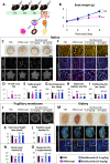
Fig 2. The effect of ranibizumab and aflibercept injected intraperitoneally into neonatal mice.
(A) A schematic diagram depicting the time course of our experiment. (B) The body weight gains were impaired in the aflibercept group but not in the ranibizumab groups. (C-E) The whole-mounted retinas showed apparent retinal hemorrhage (arrowheads) and vascular growth impairment in the aflibercept group but not in the ranibizumab groups. (F-I) The high magnification images of the whole-mounted retinas show decreased number of branching points, increased area of regressed capillaries (filled arrowheads), and decreased number of proliferating ECs (open arrowheads) in the aflibercept group but not in the ranibizumab groups. (J-L) The whole-mounted pupillary membranes showed decreased pupillary vessel length and density in the aflibercept group but not in the ranibizumab groups. (M-O) The kidney sections showed pathological renal thrombosis (arrowheads), decreased density of renal vessels, and decreased number of glomerular endothelial cells in the aflibercept group but not in the ranibizumab groups. Scale bars represent 1000 μm (C, top and middle rows of M); 500 μm (J); 100 μm (F and bottom row of M); **P<0.01; *P<0.05 (n = 6–7 /group). ECs, endothelial cells; NS, not significant; PBS, phosphate-buffered saline solution.
Fig 3
Fig 3. Western blot analysis of the interaction of recombinant human VEGF-A165, and mouse VEGF-A164, human VEGF-A121, and mouse VEGF-A120 with aflibercept or ranibizumab as the primary antibody.
The immunoreactive bands for mouse VEGF-A were observed in the aflibercept-probed blot, but not in the ranibizumab-probed blot even with long exposure time. VEGF, vascular endothelial growth factor.

References

    1. Brown DM, Kaiser PK, Michels M, Soubrane G, Heier JS, Kim RY, et al.; ANCHOR Study Group. Ranibizumab versus verteporfin for neovascular agerelated macular degeneration. N Engl J Med. 2006. Oct 5;355(14):1432–1444. - PubMed
    1. CATT Research Group, Martin DF, Maguire MG, Ying GS, Grunwald JE, Fine SL, et al.. Ranibizumab and bevacizumab for neovascular agerelated macular degeneration. N Engl J Med. 2011. May 19;364(20):1897–1908. doi: 10.1056/NEJMoa1102673 - DOI - PMC - PubMed
    1. Heier JS, Brown DM, Chong V, Korobelnik JF, Kaiser PK, Nguyen QD, et al.. Intravitreal aflibercept (VEGF trap-eye) in wet age-related macular degeneration. Ophthalmology. 2012. Dec;119(12):2537–48. doi: 10.1016/j.ophtha.2012.09.006 - DOI - PubMed
    1. Diabetic Retinopathy Clinical Research Network, Wells JA, Glassman AR, Ayala AR, Jampol LM, Aiello LP, et al.. Aflibercept, bevacizumab, or ranibizumab for diabetic macular edema. N Engl J Med. 2015. Mar 26;372(13):1193–203. doi: 10.1056/NEJMoa1414264 - DOI - PMC - PubMed
    1. Jampol LM, Glassman AR, Sun J. Evaluation and Care of Patients with Diabetic Retinopathy. N Engl J Med. 2020. Apr 23;382(17):1629–1637. doi: 10.1056/NEJMra1909637 - DOI - PubMed

Publication types

MeSH terms