. 2022 Dec 21;20(12):e3001934.
doi: 10.1371/journal.pbio.3001934. eCollection 2022 Dec.

Alternative splicing liberates a cryptic cytoplasmic isoform of mitochondrial MECR that antagonizes influenza virus

Affiliations

Alternative splicing liberates a cryptic cytoplasmic isoform of mitochondrial MECR that antagonizes influenza virus

Steven F Baker et al. PLoS Biol. .

Abstract

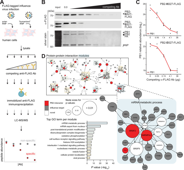
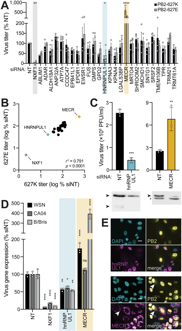
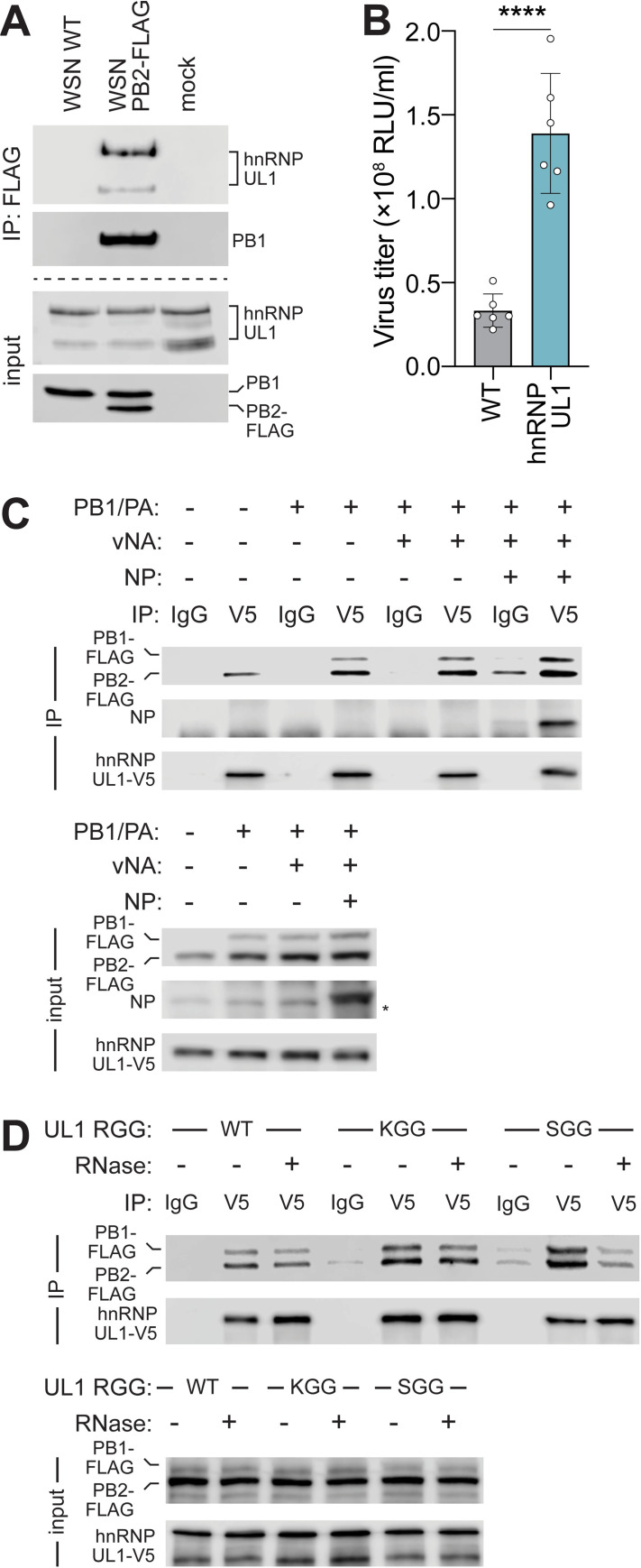
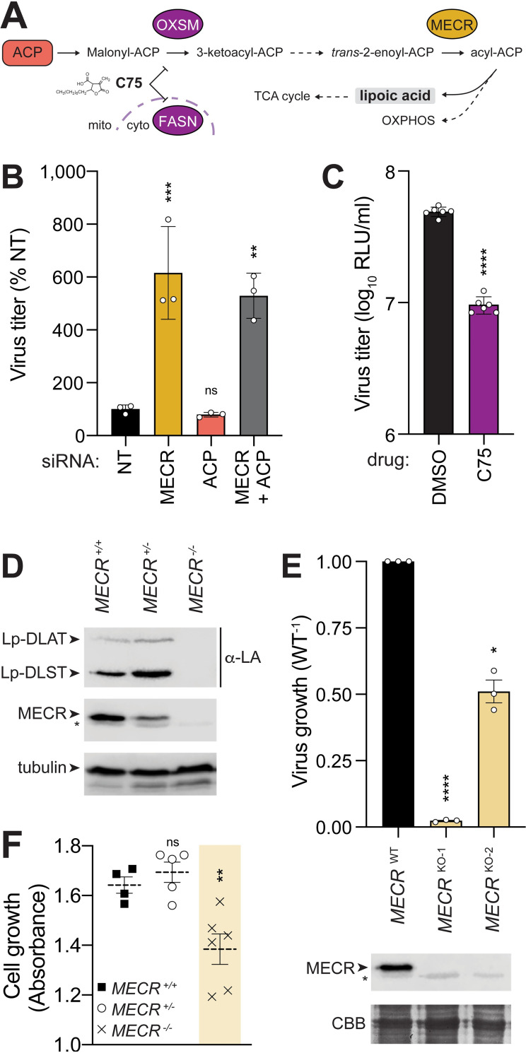
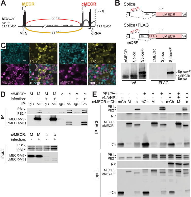
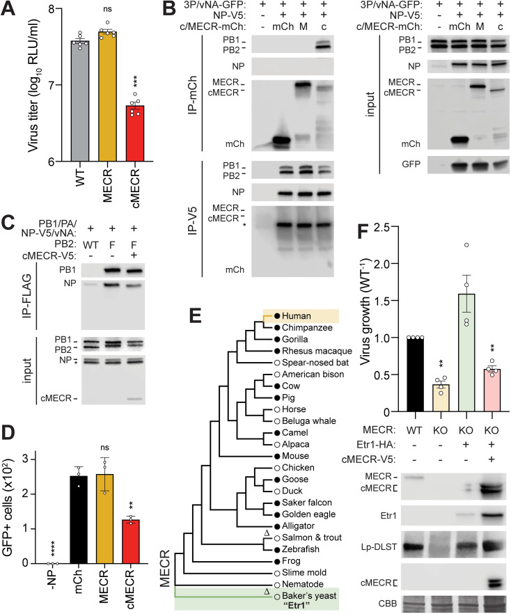
Viruses must balance their reliance on host cell machinery for replication while avoiding host defense. Influenza A viruses are zoonotic agents that frequently switch hosts, causing localized outbreaks with the potential for larger pandemics. The host range of influenza virus is limited by the need for successful interactions between the virus and cellular partners. Here we used immunocompetitive capture-mass spectrometry to identify cellular proteins that interact with human- and avian-style viral polymerases. We focused on the proviral activity of heterogenous nuclear ribonuclear protein U-like 1 (hnRNP UL1) and the antiviral activity of mitochondrial enoyl CoA-reductase (MECR). MECR is localized to mitochondria where it functions in mitochondrial fatty acid synthesis (mtFAS). While a small fraction of the polymerase subunit PB2 localizes to the mitochondria, PB2 did not interact with full-length MECR. By contrast, a minor splice variant produces cytoplasmic MECR (cMECR). Ectopic expression of cMECR shows that it binds the viral polymerase and suppresses viral replication by blocking assembly of viral ribonucleoprotein complexes (RNPs). MECR ablation through genome editing or drug treatment is detrimental for cell health, creating a generic block to virus replication. Using the yeast homolog Etr1 to supply the metabolic functions of MECR in MECR-null cells, we showed that specific antiviral activity is independent of mtFAS and is reconstituted by expressing cMECR. Thus, we propose a strategy where alternative splicing produces a cryptic antiviral protein that is embedded within a key metabolic enzyme.


Conflict of interest statement

I have read the journal’s policy and the authors of this manuscript have the following competing interests: AM is an editorial board member for PLoS Biology and PLoS Pathogens. HM, MT, SG, JSP, AA and HJ were employees of F. Hoffmann-La Roche when performing this work. No other authors declare a competing interest.

Figures

Fig 1
Fig 1. ICC-MS defines an influenza polymerase interactome.
(A) Schematic diagram to identify PB2 interactome. Lysate fractions from human A549 cells infected with PB2-FLAG-tagged influenza virus encoding avian RNP or the “humanized” PB2-E627K RNP were incubated with competing soluble anti-FLAG antibody followed by capture with resin-bound anti-FLAG and LC–MS/MS. (B-C) Immunocompetitive capture of the viral RNP. (B) Detection of PB2 627E and 627K (top: western blot) or PB2 627K with coprecipitating RNP components (bottom: silver stain). *, IgG heavy chain. (C) Relative protein abundance of PB1 in PB2 ICC-MS samples shows decreasing capture with increasing competition antibody. Data shown are in biological triplicate. (D) Minimum-cost flow simulations connect top PB2 interactors identified by ICC-MS (red) to previously identified influenza host factors (gray), in some cases through other host proteins (white). Modules comprising different PB2 interactors were enriched for GO terms, the most significant of which is enlarged. Node sizes indicate empirical P values derived from the control flow simulations. Individual quantitative observations that underlie the data summarized here can be located under the Supporting information file as S1 Data. Uncropped images can be found in the Supporting information file as S1 Raw Images. Ab, antibody; ICC-MS, immunocompetitive capture-mass spectrometry; IgG, immunoglobulin G; LC-MS/MS, liquid chromatography-tandem mass spectrometry; NP, nucleoprotein; RNP, ribonucleoprotein complex.
Fig 2
Fig 2. Functional analysis of top candidate PB2 interactors reveals important roles for hnRNP UL1 and MECR.
(A) Secondary screening of proteomic hits by siRNA treatment and reporter virus infection. After knockdown, A549 cells were infected with human (PB2-627K; MOI, 0.01) or avian-adapted (PB2-627E; MOI, 0.05) WSN NLuc virus for 24 h. Viral supernatants were titered and normalized to an NT control. Control NXF1 (gray) and outliers hnRNP UL1 (cyan) and MECR (yellow) are highlighted. (B) Concordance of virus titer for PB2-627E vs. PB2-627K virus infections in siRNA-treated cells (from (A)). Statistical analysis performed with a two-tailed Pearson correlation coefficient. (C) Multicycle virus replication of WT virus was measured 24 h postinfection in A549 cells treated with the indicated siRNAs. Knockdown efficiency was analyzed by western blot. Asterisk indicates nonspecific band. (D) Knockdown impacts viral gene expression of divergent influenza viruses. siRNA-treated A549 cells were infected with reporter viruses based on WSN (pre-2009 H1N1; MOI, 0.1), CA04 (pandemic 2009 H1N1; MOI, 0.5), or B/Bris (Victoria-lineage; MOI, 1). Viral gene expression was measured 8 h postinfection and normalized to NT controls. (E) A549 cells stably expressing hnRNP UL1 (top) or MECR (bottom) were infected with WSN PB2-FLAG (MOI 3, 8 h). Protein localization was detected by immunofluorescence, and nuclei were visualized with DAPI. Arrow indicates minor PB2 population consistent with previously reported patterns of mitochondrial localization. Scale bars, 20 μm. Data in (A) are mean ± SEM of n = 3 biological replicates. Comparisons were performed with two-way ANOVA with post hoc Fisher LSD test. For (C) and (D), data are mean ± SD of n = 3. Comparisons were performed with a two-tailed Student t test (C) or a two-way ANOVA with post hoc Dunnett multiple comparisons test (D); *, P < 0.05; **, P < 0.01; ***, P < 0.001; ****, P < 0.0001; ns, not significant. Individual quantitative observations that underlie the data summarized here can be located under the Supporting information file as S1 Data. Uncropped images can be found in the Supporting information file as S1 Raw Images. hnRNP UL1, heterogenous nuclear ribonuclear protein U-like 1; MECR, mitochondrial enoyl CoA-reductase; NLuc, nanoluciferase; NT, nontargeting; WT, wild-type.
Fig 3
Fig 3. Proviral hnRNP UL1 associates with influenza polymerase.
(A) Endogenous hnRNP UL1 coprecipitates with PB2 during infection. A549 cells were infected (MOI, 1; 18 h) or mock treated, lysed, and immunoprecipitated. Proteins were detected by western blot. (B) Viral titers were measured from WT or clonal A549 hnRNP UL1-V5 cells infected with WSN Nluc (MOI, 0.05; 24 h). Mean ± SD of n = 6. Unpaired two-tailed t test; ****, P < 0.0001. (C-D) Association of viral polymerase with hnRNP UL1. (C) hnRNP UL1-V5 was immunoprecipitated with anti-V5 antibody or control IgG from lysates generated from cells expressing the indicated viral proteins or RNA (vNA).Proteins were detected by western blotting. Asterisk indicates nonspecific band. (D) Co-immunoprecipitation assays were performed using WT hnRNP UL1 or mutants disrupted in methylation (KGG) or RNA binding (SGG). RNase was included during immunoprecipitation where indicated. Individual quantitative observations that underlie the data summarized here can be located under the Supporting information file as S1 Data. Uncropped images can be found in the Supporting information file as S1 Raw Images. hnRNP UL1, heterogenous nuclear ribonuclear protein U-like 1; IgG, immunoglobulin G; IP, immunoprecipitation; NLuc, nanoluciferase; NP, nucleoprotein; WT, wild-type.
Fig 4
Fig 4. Modulating the critical mtFAS enzyme MECR alters mtFAS output and virus growth.
(A) Focused snapshot of the mtFAS pathway. ACP and MECR were experimentally probed by knockdown or knockout, whereas OXSM and FASN were inhibited with the drug C75. (B) A549 cells were treated with siRNA targeting MECR, ACP, both, or an NT control prior to infection with WSN NLuc virus (MOI, 0.05; 24 h). Viral titer in supernatants was determined and normalized to NT. Mean ± SD of n = 3. One-way ANOVA with post hoc Dunnett multiple comparisons test; **, P < 0.01; ***, P < 0.001; ns, not significant. (C) Viral yield was measured from A549 cells treated with C75 or DMSO control prior to infection with WSN NLuc (MOI, 0.05; 24 h). Mean ± SD of n = 6. Unpaired two-tailed t test; ****, P < 0.0001. (D) Production of lipoylated subunits of the pyruvate dehydrogenase complex (DLAT) and the 2-oxoglutarate dehydrogenase complex (DLST) was assessed in WT (+/+), heterozygous (+/−), and homozygous (−/−) MECR knockout A549 cells by western blotting with anti-lipoic acid (α-LA) antibody. (E) Virus replication was measured in A549 cells (MOI, 0.05; 24 h). Replication in MECR knockout clones KO-1 and KO-2 was normalized to WT A549 cells. Mean ± SEM of biological replicates (n = 3) normalized to WT. MECR expression was monitored by western blotting. CBB staining was used as a loading control. Asterisks indicate nonspecific bands. (F) Growth of clonal WT, heterozygous, or homozygous MECR knockout A549 cells was measured over 3 d. Mean ± SEM of n = 4–6 clones. One-way ANOVA with post hoc Dunnett multiple comparisons test; **, P < 0.01; ns, not significant. Individual quantitative observations that underlie the data summarized here can be located under the Supporting information file as S1 Data. Uncropped images can be found in the Supporting information file as S1 Raw Images. ACP, acyl carrier protein; CBB, Coomassie brilliant blue; DLAT, dihydrolipoamide acetyltransferase; DLST, dihydrolipoamide S-succinyltransferase; FASN, fatty acid synthase; MECR, mitochondrial enoyl CoA-reductase; mtFAS, mitochondrial fatty acid synthesis; NLuc, nanoluciferase; NT, nontargeting; OXSM, 3-oxoacyl-ACP synthase, mitochondrial; WT, wild-type.
Fig 5
Fig 5. The alternative splice variant cMECR binds viral polymerase subunit PB2.
(A) Sashimi plot of RNA-seq data from A549 cells showing alternative 3′ splice site utilized by transcripts that do not translate the MTS in exon 1. Numbers embedded in yellow (MECR) and red (cMECR) curves indicate percentage of each splicing event as a total of all exon-joining reads. Arrows indicate translational start sites and gRNA denotes region targeted in CRISPR-Cas9 editing. (B) Splicing reporters were constructed as diagrammed and expressed in 293T cells with cMECR cDNA as a control. Proteins were detected by western blot. (C) 293T cells expressing MECR or cMECR were infected with influenza PB2-FLAG virus (MOI, 3; 8 h). Subcellular localization of PB2, MECR, or cMECR were determined by immunofluorescence microscopy. Scale bars, 20 μm. (D-E) The viral polymerase subunit PB2 associates with cMECR during infection. (D) PA, PB1-FLAG, PB2-FLAG, and V5-tagged MECR or cMECR were expressed in 293T cells. Where indicated, cells were also infected with WSN (MOI, 10; 6 h). Cells were lysed and immunoprecipitated with anti-V5 antibody or IgG controls. Proteins were detected by western blot. (E) mCh, MECR M77L-mCh, or cMECR-mCh were coexpressed with PB2-FLAG in the absence or presence of PB1-FLAG, PA, NP, and vNA. Cells were lysed and immunoprecipitated with mCh affinity resin. Proteins were detected by western blot. Uncropped images can be found in the Supporting information file as S1 Raw Images. cMECR, cytoplasmic MECR; IgG, immunoglobulin G; IP, immunoprecipitation; mCh, mCherry; MECR, mitochondrial enoyl CoA-reductase; MTS, mitochondrial targeting signal; NP, nucleoprotein; vNA, NA viral RNA.
Fig 6
Fig 6. The antiviral activity of cMECR inhibits RNP assembly independent of MECR and mtFAS.
(A) Replication of WSN NLuc was measured in WT or clonal A549 cells expressing MECR or cMECR. Protein expression from infected cells was analyzed by western blotting. Asterisk indicates a nonspecific band used as a loading control. Mean ± SD of n = 6. One-way ANOVA with post hoc Dunnett multiple comparisons test; ***, P < 0.001; ns, not significant. (B-C) cMECR disrupts RNP assembly. (B) RNPs were assembled in 293T cells coexpressing mCh, MECR M77L-mCh, or cMECR-mCh. NP was omitted in the negative control (−NP). Cells were lysed, divided in half, and immunoprecipitated for MECR (mCh) or NP (V5). Input or coprecipitated proteins were detected by western blot. * = NP detected from prior probing of the membrane before blotting for mCh. (C) RNP assembly monitored as in (B) except PB2 (FLAG) was targeted for immunoprecipitation with untagged PB2 as the negative control. (D) Polymerase activity assays with vNA-GFP were performed in the presence of mCh, MECR M77L-mCh, or cMECR-mCh. Fluorescence microscopy images taken at 24 h posttransfection and GFP-positive cells were enumerated by ImageJ. Mean ± SD of n = 3. Two-way ANOVA with post hoc Dunnett multiple comparisons test; ***, P < 0.001; **, P < 0.01; ns, not significant. (E) Phylogenetic maximum-likelihood analysis of MECR amino acid sequences. Δ, sequences lacking conserved cMECR start codon (Met77, human numbering). Circles indicate presence (filled circles) or absence (empty circles) of annotated cMECR transcripts. NCBI RefSeq IDs listed in S4 Table. (F) MECR KO cells were complemented with the MECR homolog from Saccharomyces cerevisiae Etr1 then transduced with cMECR. Cells were subsequently infected with WSN NLuc (MOI, 0.05; 24 h), and viral titers were measured in supernatants and compared to WT. Mean ± SD of n = 4. Unpaired two-tailed t test; *, P < 0.05; **, P < 0.01. Below, mtFAS rescue was confirmed by detecting Lp-DLAT via western blotting whole cell lysates. Individual quantitative observations that underlie the data summarized here can be located under the Supporting information file as S1 Data. Uncropped images can be found in the Supporting information file as S1 Raw Images. CBB, Coomassie brilliant blue; cMECR, cytoplasmic MECR; Lp-DLAT, lipoylated dihydrolipoamide acetyltransferase; Lp-DLST, lipoylated dihydrolipoamide S-succinyltransferase; KO, knockout; mCh, mCherry; MECR, mitochondrial enoyl CoA-reductase; mtFAS, mitochondrial fatty acid synthesis; NLuc, nanoluciferase; NP, nucleoprotein; RNP, ribonucleoprotein complex; WT, wild-type.

References

    1. Garten R, Blanton L, Elal AIA, Alabi N, Barnes J, Biggerstaff M, et al.. Update: Influenza Activity in the United States During the 2017–18 Season and Composition of the 2018–19 Influenza Vaccine. MMWR Morbidity and Mortality Weekly Report. 2018;67:634–642. doi: 10.15585/mmwr.mm6722a4 - DOI - PMC - PubMed
    1. Van Valen L. The Red Queen lives. Nature. 1976;260:575. doi: 10.1038/260575a0 - DOI
    1. Daugherty MD, Malik HS. Rules of engagement: Molecular insights from host-virus arms races. Annual Review of Genetics. 2012;46:677–700. doi: 10.1146/annurev-genet-110711-155522 - DOI - PubMed
    1. Rialdi A, Hultquist J, Jimenez-Morales D, Peralta Z, Campisi L, Fenouil R, et al.. The RNA Exosome Syncs IAV-RNAPII Transcription to Promote Viral Ribogenesis and Infectivity. Cell. 2017;169:679–692.e14. doi: 10.1016/j.cell.2017.04.021 - DOI - PMC - PubMed
    1. Sugiyama K, Kawaguchi A, Okuwaki M, Nagata K. PP32 and APRIL are host cell-derived regulators of influenza virus RNA synthesis from cRNA. eLife. 2015;4:e08939. doi: 10.7554/eLife.08939 - DOI - PMC - PubMed

Publication types