. 2022 Dec 21;14(676):eade0424.
doi: 10.1126/scitranslmed.ade0424. Epub 2022 Dec 21.

A prefusion-stabilized RSV F subunit vaccine elicits B cell responses with greater breadth and potency than a postfusion F vaccine

Affiliations

A prefusion-stabilized RSV F subunit vaccine elicits B cell responses with greater breadth and potency than a postfusion F vaccine

Lauren A Chang et al. Sci Transl Med. .

Abstract

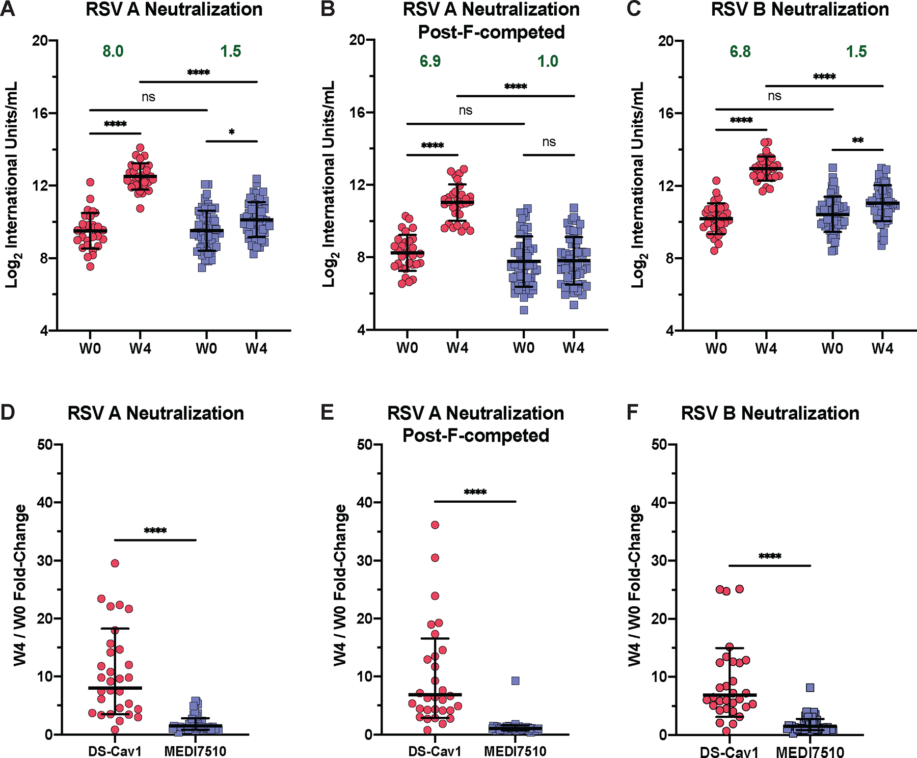
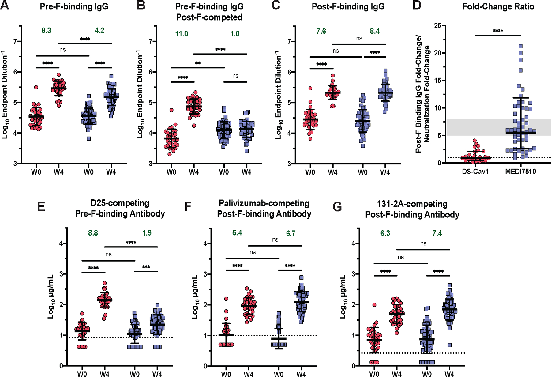
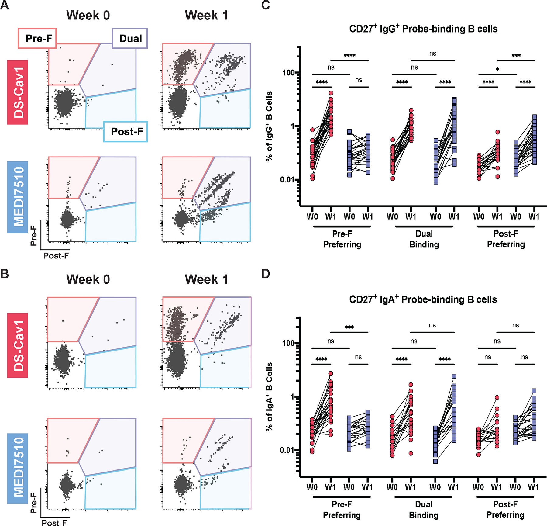
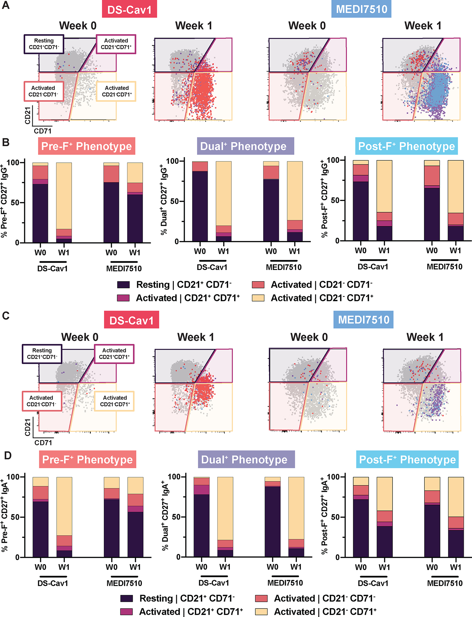
There is currently no licensed vaccine for respiratory syncytial virus (RSV). Here, we assess the effect of RSV fusion protein (F) conformation on B cell responses in a post hoc comparison of samples from the DS-Cav1 [prefusion (pre-F)] and MEDI7510 [postfusion (post-F)] vaccine clinical trials. We compared the magnitude and quality of the serological and B cell responses across time points and vaccines. We measured RSV A and B neutralization, F-binding immunoglobulin G titers, and competition assays at week 0 (before vaccination) and week 4 (after vaccination) to evaluate antibody specificity and potency. To compare B cell specificity and activation, we used pre-F and post-F probes in tandem with a 17-color immunophenotyping flow cytometry panel at week 0 (before vaccination) and week 1 (after vaccination). Our data demonstrate that both DS-Cav1 and MEDI7510 vaccination robustly elicit F-specific antibodies and B cells, but DS-Cav1 elicited antibodies that more potently neutralized both RSV A and B. The superior potency was mediated by antibodies that bind antigenic sites on the apex of pre-F that are not present on post-F. In the memory (CD27+) B cell compartment, vaccination with DS-Cav1 or MEDI7510 elicited B cells with different epitope specificities. B cells preferentially binding the pre-F probe were activated in DS-Cav1-vaccinated participants but not in MEDI7510-vaccinated participants. Our findings emphasize the importance of using pre-F as an immunogen in humans because of its deterministic role in eliciting highly potent neutralizing antibodies and memory B cells.

Trial registration: ClinicalTrials.gov NCT02508194 NCT02289820 NCT03049488.


Conflict of interest statement

Competing interests: B.S.G. is on the scientific advisory board for Icosavax, Greenlight Biosciences, Vaccine Company, Inc, Foundry, and Afrigen WHO mRNA Biohub and is an ad hoc consultant for GSK, Pfizer, Janssen, AstraZeneca, Merck, and Sanofi. He is an inventor on patents for the stabilization of the RSV F protein (WO2014160463A1, Prefusion RSV F proteins and their use). M.T.E., T.V., J.F., and F.D. were all employees at AstraZeneca when the MEDI7510 clinical trial was performed and may own stock in AstraZeneca. All other authors declare no competing interests.

Figures

Figure 1.
Figure 1.. Pre-F vaccination elicits a greater magnitude of pre-F-exclusive and highly neutralizing antibodies than post-F vaccination.
(A to C) Conformation-specific neutralization capacity of serum obtained at week 0 (W0) and week 4 (W4) after vaccination with DS-Cav1 (pre-F, N = 30) or MEDI7510 (post-F, N = 52) was measured for (A) RSV subtype A2 without post-F competition or (B) with post-F competition, and (C) subtype B. In (B), excess post-F was used to adsorb post-F-exclusive and dual-binding antibodies to assess the neutralization capacity of pre-F exclusive antibodies in serum. (D to F) Fold-change in neutralization capacity at W4 after vaccination with either DS-Cav1 or MEDI7510 was calculated for RSV subtype A2 (D) without or (E) with post-F competition, and (F) subtype B. Bars represent mean ± standard deviation (SD) of log2 data. Dotted lines indicate the limit of detection (LOD) for each assay. For all graphs, asterisks denote the P-value, with ****P < 0.0001, ***P = 0.0001 to 0.001, **P = 0.001 to 0.01, *P = 0.01 to 0.05, and ns = not significant as determined using Brown-Forsythe and Welch ANOVA tests (A to C), Kruskal-Wallis tests (D and E), or Mann-Whitney tests (F). Red circles denote participants who received DS-Cav1, blue squares denote participants who received MEDI7510. Geometric mean W4/W0 fold-change is shown in green text.
Figure 2.
Figure 2.. Pre-F vaccination elicits pre-F-exclusive binding antibody and higher neutralization potency.
(A) W0 and W4 pre-F binding IgG endpoint titers are shown for serum samples from participants after vaccination with DS-Cav1 (pre-F, N = 30) or MEDI7510 (post-F, N = 52). (B) Pre-F binding IgG endpoint titers are shown as in (A), except excess post-F was used to adsorb post-F-exclusive and dual-binding antibodies from serum to leave pre-F-exclusive binding antibodies. W0 and W4 (C) post-F binding IgG endpoint titers are shown. (D) The fold-change ratio of post-F binding to neutralization is shown for each participant. Fold-change ratio = (W4/W0 fold-change, post-F IgG binding)/(W4/W0 fold-change, RSV A neutralization). A ratio below 1 indicates the increase in neutralization capacity exceeded the increase in binding capacity, demonstrating the elicitation of highly potent neutralizing antibodies. The gray area represents the historical range of this ratio observed in previous clinical trials of post-F subunit vaccines (fold-change ratio = 5 to 8). (E to G) Concentrations of (E) pre-F-binding D25-competing antibodies (DCA), (F) post-F-binding palivizumab-competing antibodies (PCA), and (G) post-F-binding 131–2A-competing antibodies (1CA) in serum are shown for samples collected before (W0) or 4 weeks after (W4) vaccination with DS-Cav1 or MEDI7510. Bars represent mean ± SD of log10 data. Dotted lines indicate the lower limit of quantitation (LLOQ) for (E to G), with undetectable values arbitrarily set to ½ LLOQ. Fold-change and statistics were calculated using values above the LLOQ. For all graphs, asterisks denote the P-value, with ****P < 0.0001, ***P = 0.0001 to 0.001, **P = 0.001 to 0.01, *P = 0.01 to 0.05, and ns = not significant as determined using Brown-Forsythe and Welch ANOVA tests (A to C, G), Mann-Whitney tests (D), or Kruskal-Wallis tests (E and F). Red circles denote participants who received DS-Cav1, blue squares denote participants who received MEDI7510. Geometric mean W4/W0 fold-change is shown in green text.
Figure 3.
Figure 3.. Pre-F vaccination elicits pre-F-preferring and dual-binding memory B cells.
Representative pre-F and post-F probe-binding profiles from a single participant vaccinated with DS-Cav1 (pre-F, N = 30) or MEDI7510 (post-F, N = 30) at W0 and W1 are shown. Fluorescently labeled pre-F or post-F probes were used to separate class-switched memory B cells into three compartments (pre-F-preferring, dual-binding, or post-F-preferring). IgG+ (A) representative binding profile and (B) quantification of probe-binding B cells are shown. IgA+ (C) representative binding profile and (D) quantification of probe-binding B cells are shown. For all graphs, asterisks denote the P-value, with ****P < 0.0001, ***P = 0.0001 to 0.001, **P = 0.001 to 0.01, *P = 0.01 to 0.05, and ns = not significant as determined using Brown-Forsythe and Welch ANOVA tests (B) or Kruskal-Wallis tests (D). Red circles denote participants who received DS-Cav1, blue squares denote participants who received MEDI7510.
Figure 4.
Figure 4.. Activation phenotypes of memory B cells following pre-F or post-F vaccination parallels F probe-binding profiles.
Representative pre- and post-F probe-binding profiles are shown for a single participant vaccinated with DS-Cav1 (pre-F, N = 30) or MEDI7510 (post-F, N = 30) at W0 and W1. CD27+ B cells were delineated based on expression of surface markers CD27 and CD21 as activated (CD21+CD71+, CD21CD71, CD21CD71+) or resting (CD21+CD71). F probe-binding B cells were overlaid for each compartment. IgG+ (A) representative activation profiles and (B) activation phenotype by F probe-binding specificity are shown. IgA+ (C) representative activation profiles and (D) activation phenotype by F probe-binding specificity are shown.

Comment in

References

    1. Aiello A et al., Immunosenescence and Its Hallmarks: How to Oppose Aging Strategically? A Review of Potential Options for Therapeutic Intervention. Front. Immunol. 10, 2247 (2019). - PMC - PubMed
    1. Beem M, Wright FH, Hamre D, Egerer R, Oehme M, Association of the chimpanzee coryza agent with acute respiratory disease in children. N. Engl. J. Med. 263, 523–530 (1960). - PubMed
    1. Hall CB, Simőes EAF, Anderson LJ, in Challenges and Opportunities for Respiratory Syncytial Virus Vaccines, Anderson LJ, Graham BS, Eds. (Springer Berlin Heidelberg, Berlin, Heidelberg, 2013), pp. 39–57.
    1. Hall CB, The burgeoning burden of respiratory syncytial virus among children. Infect. Disord. Drug Targets 12, 92–97 (2012). - PubMed
    1. Shi T et al., Global, regional, and national disease burden estimates of acute lower respiratory infections due to respiratory syncytial virus in young children in 2015: a systematic review and modelling study. Lancet 390, 946–958 (2017). - PMC - PubMed

Publication types

MeSH terms

Associated data