. 2022 Dec 21;14(676):eadd0484.
doi: 10.1126/scitranslmed.add0484. Epub 2022 Dec 21.

Persistent post-COVID-19 smell loss is associated with immune cell infiltration and altered gene expression in olfactory epithelium

Affiliations

Persistent post-COVID-19 smell loss is associated with immune cell infiltration and altered gene expression in olfactory epithelium

John B Finlay et al. Sci Transl Med. .

Abstract

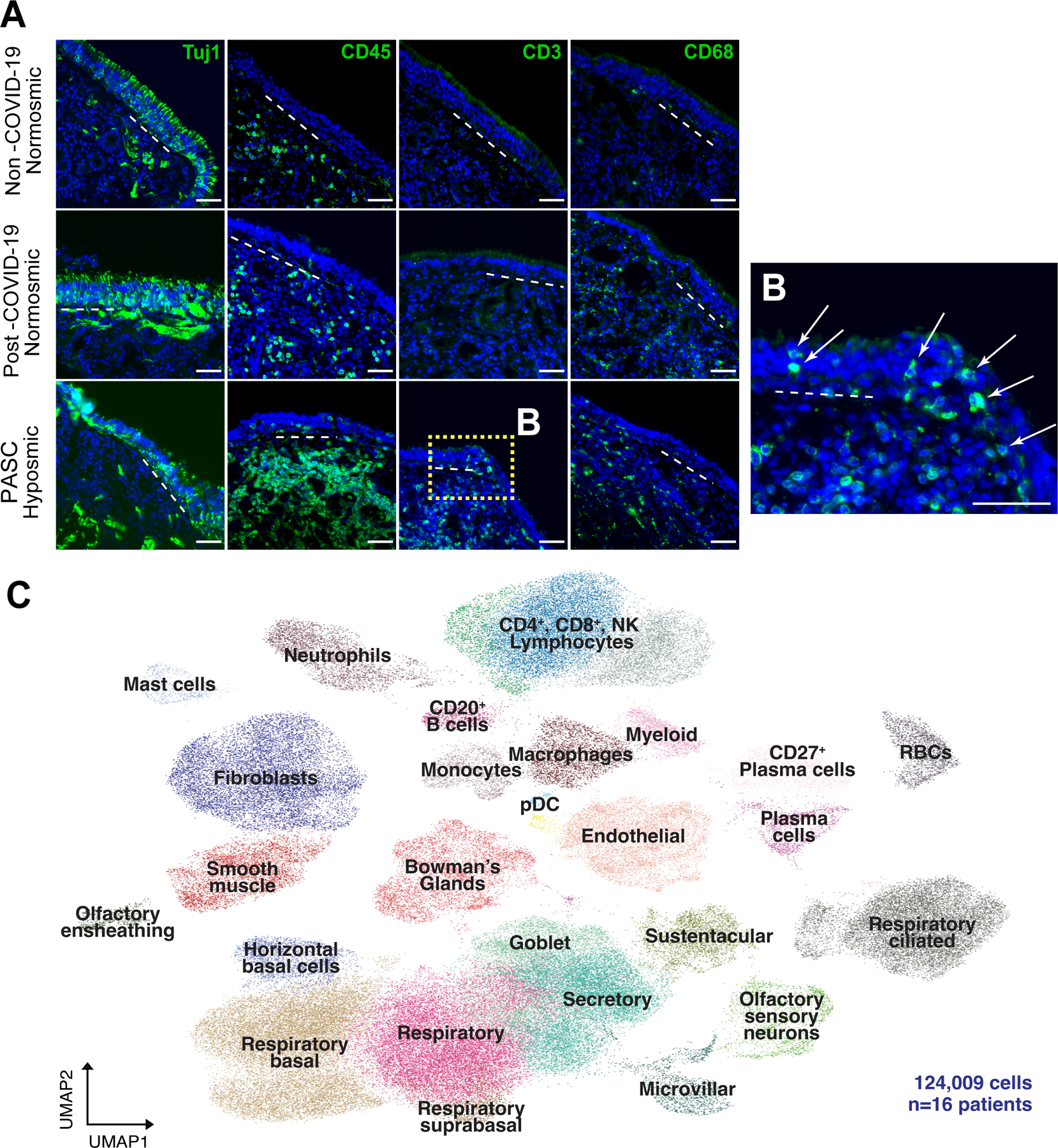
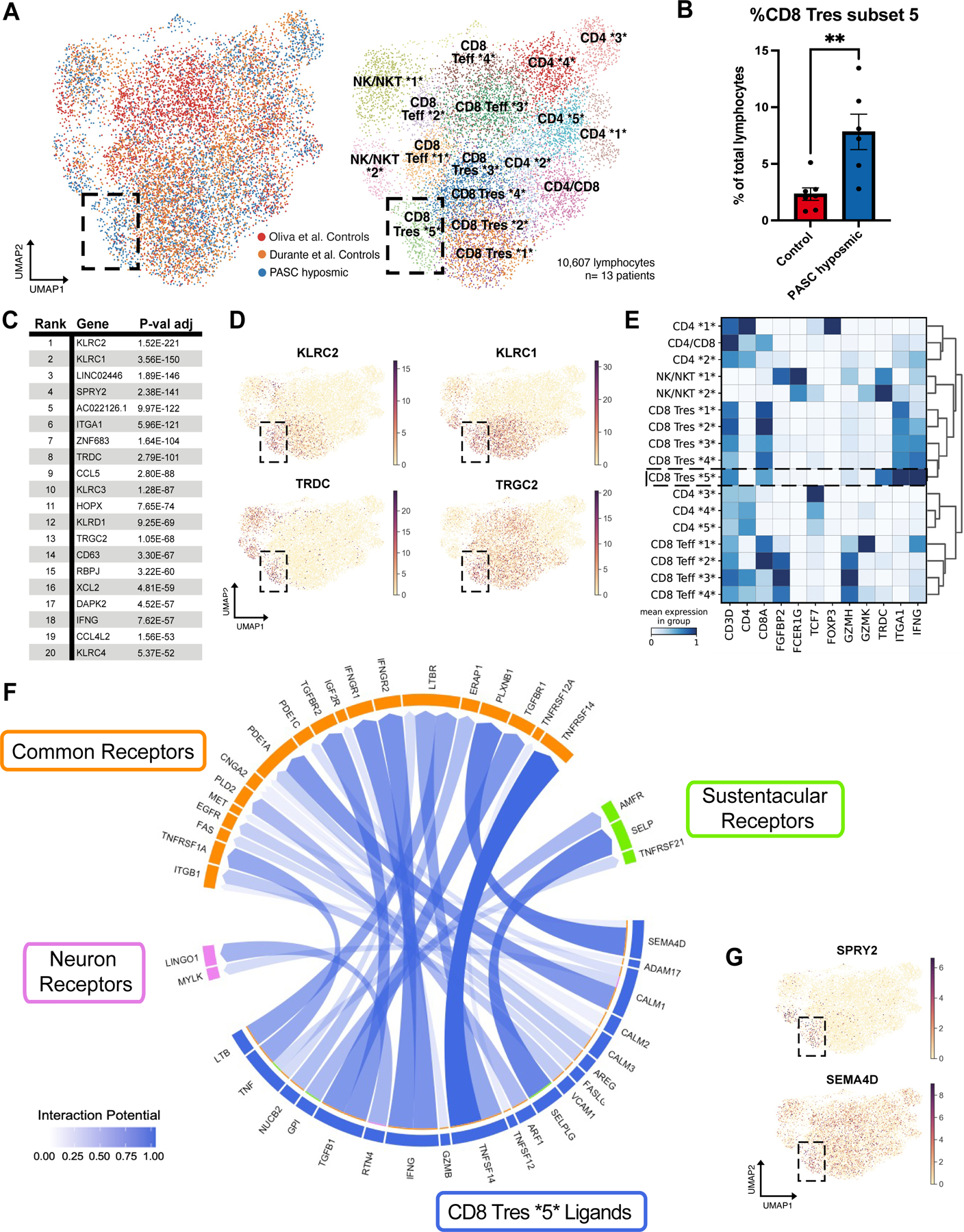
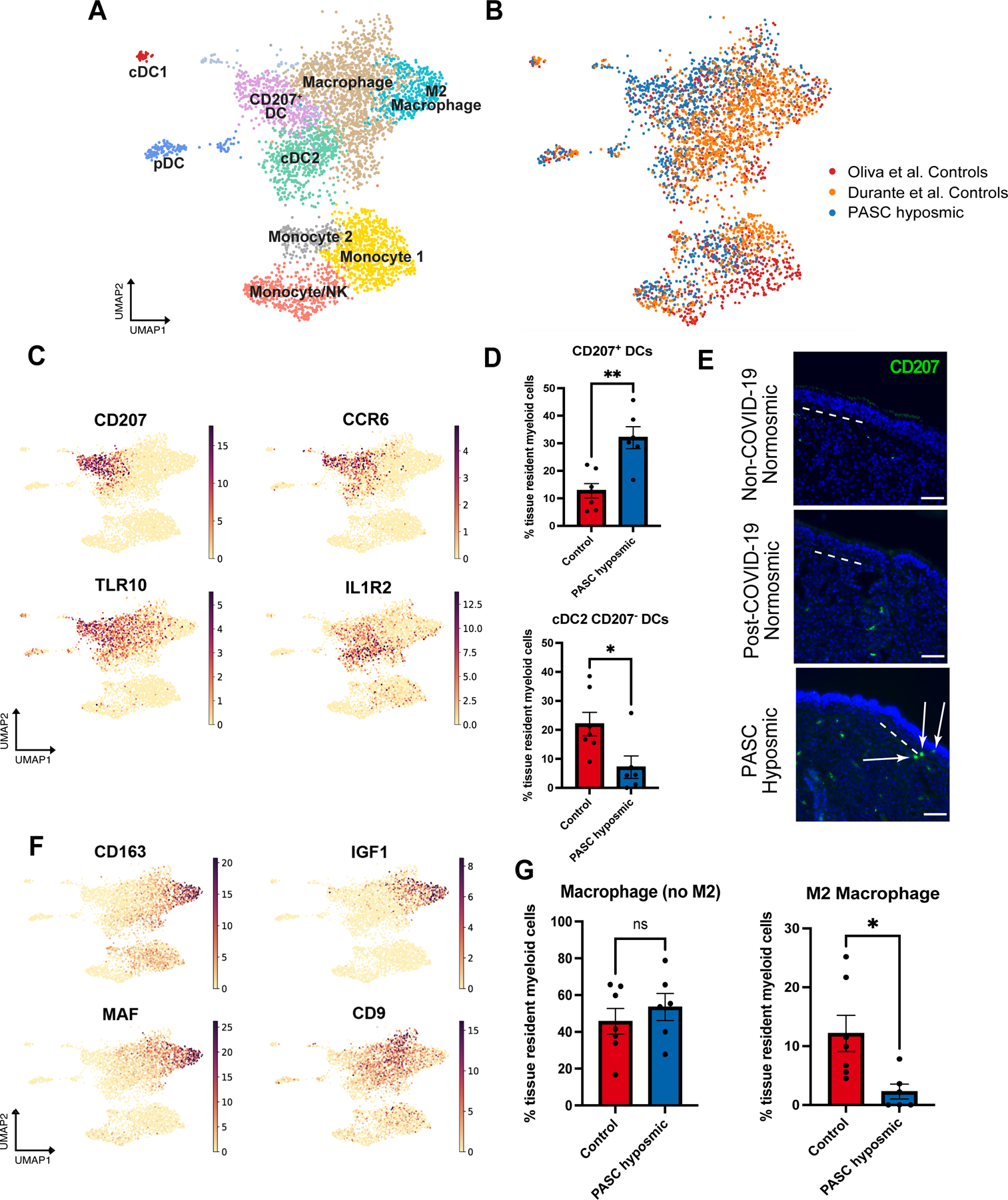
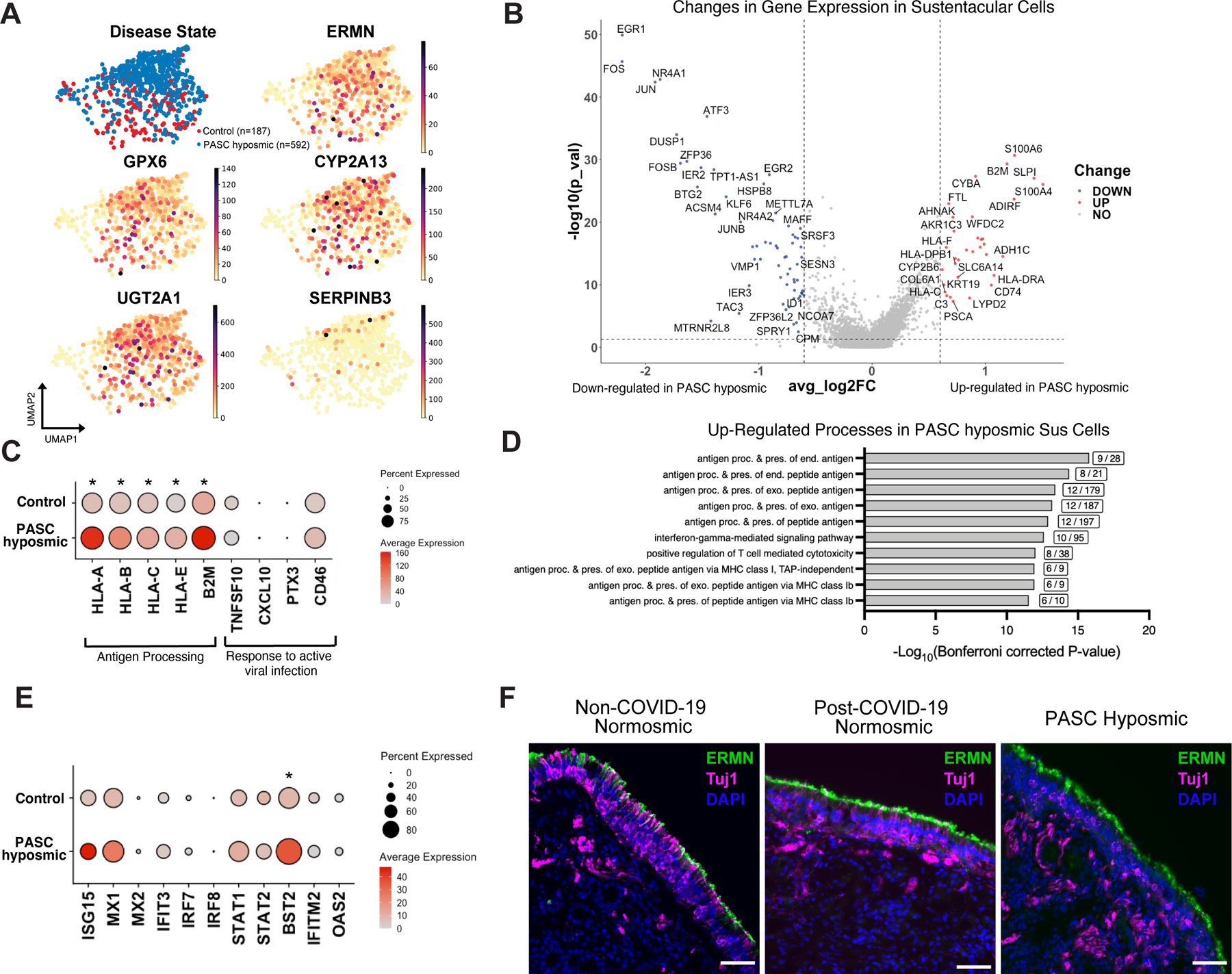
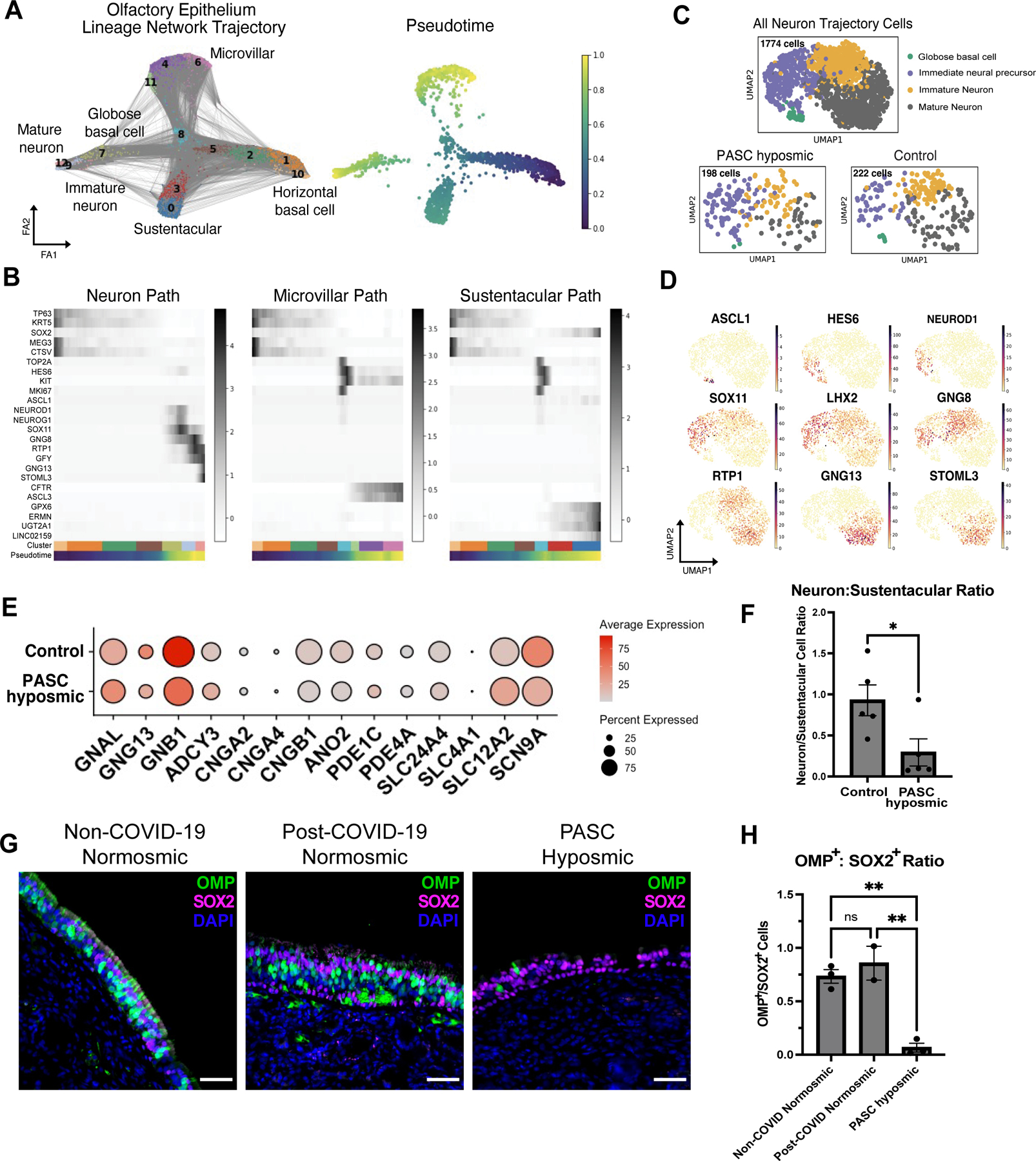
SARS-CoV-2 causes profound changes in the sense of smell, including total smell loss. Although these alterations are often transient, many patients with COVID-19 exhibit olfactory dysfunction that lasts months to years. Although animal and human autopsy studies have suggested mechanisms driving acute anosmia, it remains unclear how SARS-CoV-2 causes persistent smell loss in a subset of patients. To address this question, we analyzed olfactory epithelial samples collected from 24 biopsies, including from nine patients with objectively quantified long-term smell loss after COVID-19. This biopsy-based approach revealed a diffuse infiltrate of T cells expressing interferon-γ and a shift in myeloid cell population composition, including enrichment of CD207+ dendritic cells and depletion of anti-inflammatory M2 macrophages. Despite the absence of detectable SARS-CoV-2 RNA or protein, gene expression in the barrier supporting cells of the olfactory epithelium, termed sustentacular cells, appeared to reflect a response to ongoing inflammatory signaling, which was accompanied by a reduction in the number of olfactory sensory neurons relative to olfactory epithelial sustentacular cells. These findings indicate that T cell-mediated inflammation persists in the olfactory epithelium long after SARS-CoV-2 has been eliminated from the tissue, suggesting a mechanism for long-term post-COVID-19 smell loss.


Conflict of interest statement

Competing interests: B.J.G. has received consultant fees from Frequency Therapeutics and discloses unpaid consulting to Rhino Therapeutics. D.J. has received research support from Medtronic and Association for Migraine Disorders. H.M. has received royalties from Chemcom, has received research grants from Givaudan and has received consultant fees from Kao. Authors declare no other competing interests.

Figures

Fig. 1.
Fig. 1.. T cell infiltrates in olfactory epithelium of nasal biopsies from PASC hyposmic patients.
(A) Shown are representative immunohistochemistry images of nasal biopsy tissue from normosmic non-COVID-19, normosmic post-COVID-19 or PASC hyposmic individuals. Tissue sections were immunostained for the TUJ1 neuronal marker, CD45 pan-immune cell marker, CD3 T cell marker and CD68 myeloid cell marker. PASC hyposmic tissue showed dense CD45+ immune cell infiltration including prominent CD3+ lymphocytic infiltration, which was absent in the normosmic groups; scattered CD68+ cells were present in all conditions. (B) Enlarged area (dashed yellow box) shows CD3+ lymphocytes, with prominent epithelial infiltration (arrows); dashed white lines mark the basal lamina. Scale bar, 50 μm. (C) Because of these observations, additional biopsies were processed for scRNA-seq to permit quantitative analyses; uniform manifold approximation projection (UMAP) visualization of combined PASC hyposmic and control normosmic scRNA-seq datasets integrating n=16 human nasal biopsies permitted robust cell cluster analysis and annotation. Red blood cells (RBCs), plasmacytoid dendritic cell (pDC).
Fig. 2.
Fig. 2.. A CD8+ T cell subset is enriched in PASC hyposmic nasal biopsies.
(A) UMAP visualization of all lymphocytes from CD8+, CD4+, and NK/NKT cell clusters from olfactory biopsy scRNA-seq data, comparing PASC hyposmic samples to 2 available normosmic control samples; “Oliva et al Controls” (19) were normosmic by Smell Identification Test scores, while the “Durante et al Controls” (9) were normosmic by subjective report. Black box denotes clusters enriched in PASC hyposmic samples. Teff, effector T cells; Tres, resident T cells; subsets within categories are designated by numbers (i.e. CD4 *1* thru CD4 *5*). (B) PASC hyposmic biopsies show enrichment for cell cluster CD8 Tres subset 5, compared to both control datasets (two-tailed t-test, P=0.0015). (C) Top ranked transcripts enriched in CD8 Tres subset 5 cluster (by adjusted P-value, Wilcoxon Rank-sum test, with Bonferroni correction). (D) Selected gene expression plots of significantly enriched genes in the PASC hyposmic-specific cluster CD8 Tres subset 5 (black box), including γδ T cell markers and associated genes (TRDC, TRGC2, KLRC1, KLRC2). (E) Selected gene expression and dendrogram clustering among lymphocyte subsets confirming annotations based on published marker genes. The IFNG gene is enriched in Tres subsets, especially the CD8 Tres subset 5. (F) Circos plot showing NicheNet analysis of CD8 Tres-derived ligands and their receptors in either sustentacular cells or olfactory sensory neurons, depicting interaction potential. Common receptors in orange are present in both sustentacular cells and neurons. (G) Additional plots confirm PASC hyposmic γδ T cells express T cell ligands identified by differential gene expression (SPRY2) or NicheNet analysis (SEMA4D).
Fig. 3.
Fig. 3.. PASC hyposmic olfactory samples show a myeloid cell shift with enriched dendritic cell subsets and decreased M2 macrophages.
(A) UMAP visualization of olfactory biopsy scRNA-seq data for selected myeloid cell clusters, comparing PASC hyposmic to both normosmic control data sets. DC, dendritic cells; pDC, plasmacytoid dendritic cells (CLEC4C+); cDC1, conventional DC type 1 cells (CLEC9A+); cDC2, conventional DC type 2 cells (CLEC10A+). (B) UMAP plots in panel A, colored by sample contribution. (C) Selected gene expression plots confirm cluster annotation for CD207+ dendritic cells and additional marker genes obtained by differential gene expression analysis for the CD207+ dendritic cell cluster (CCR6 and TLR10) and the cDC2 cluster (IL1R2). (D) CD207+ dendritic cells (DCs) are enriched in PASC hyposmic biopsies (blue) compared to normosmic control (non-COVID-19) biopsies (red) (two-tailed t-test, P=0.0016; CD207 DCs are reduced, P=0.0236). (E) Anti-CD207 antibody staining confirmed CD207+ dendritic cells in PASC hyposmic nasal biopsies (white arrows); dashed white lines mark the basal lamina. Scale bar, 50 μm. (F) Gene expression plots indicate marker genes for M2 macrophage (CD163, MAF, IGF1) and total macrophage (CD9) clusters. (G) PASC hyposmic biopsies showed depletion of M2 macrophages relative to all resident myeloid cells (two-tailed t-test, P=0.0175).
Fig. 4.
Fig. 4.. Sustentacular cell gene expression changes persist in PASC hyposmic olfactory epithelium.
(A) UMAP visualization of sustentacular cell subset from olfactory biopsy scRNA-seq data sets including cells from PASC hyposmic (blue) and non-COVID-19 control normosomic (red) samples (n=9 biopsies; 6 PASC hyposmic, 3 non-COVID-19 normosmic). Gene expression plots show expression of selected canonical sustentacular cell markers (ERMN, GPX6, CYP2A13) and minimal expression of the respiratory marker SERPINB3. (B) Volcano plot showing differential gene expression in sustentacular cells in PASC hyposmic compared to control normosmic olfactory epithelium biopsies; red or blue indicates significant change >0.6 log2 fold change, P<0.05). (C) Visualization of expression of selected antigen presentation genes and genes normally involved in responses to active viral infection in sustentacular cells in PASC hyposmic and control normosmic (non-COVID-19) samples (all antigen presentation genes were significantly upregulated in PASC hyposmic samples, with adjusted P value *<0.05 by Wilcoxon Rank-Sum). (D) Gene set enrichment analysis of the transcripts upregulated in PASC hyposmic sustentacular cells in (C) identifies a variety of biological processes, including “antigen presentation” and “interferon gamma signaling” (boxed figures indicate number of altered genes per process term; only significant processes are included, based on −log10(Bonferroni corrected P value)); Sus, sustentacular cell. (E) Differential gene expression of pathogen response genes previously identified in sustentacular cells in hamsters 31 days after SARS-CoV-2 infection or in uninfected hamsters (21) (BST2, an interferon-induced host gene encoding a transmembrane protein with antiviral and inflammatory signaling activities, adjusted P value <0.05; STAT1, a transcription factor downstream of inflammatory signaling, adjusted P value <0.1, Wilcoxon Rank-Sum; see also fig. S5; *=P<0.05). (F) Immunohistochemistry depicting representative labeling comparing the sustentacular cell marker ERMN (green) and the neuronal marker TUJ1 (magenta) in normosmic (non-COVID-19) control biopsies and PASC hyposmic nasal biopsies. Nuclei are stained with DAPI (blue). Scale bar, 50 μm.
Fig. 5.
Fig. 5.. Analysis of olfactory sensory neurons in PASC hyposmic samples.
(A) Trajectory lineage analysis of cells in olfactory epithelium in PASC hyposmic biopsies (n=4) from COVID-19 patients compared to control normosmic (non-COVID-19) biopsies (n=3). Cells include horizontal basal cells, globose basal cells, olfactory sensory neurons, sustentacular cells, microvillar cells. (B) Heatmaps showing pseudotime progression for olfactory epithelial cell lineages, labeled based on UMAP relations in panel A. Representative transcript markers for each cellular differentiation state are shown on the y-axis. (C) UMAP showing annotations within the olfactory sensory neuron cluster from Fig. 1D. “All Neuron Trajectory Cells” includes neuron lineage cells in biopsies from 16 patients; “PASC hyposmic” includes 5 PASC hyposmic biopsies; “Control” includes 3 control normosmic biopsies confirmed by Smell Identification Test. (D) Gene expression plots showing established markers of the olfactory sensory neuron differentiation pathway. (E) Selected gene expression in PASC hyposmic versus control normosmic biopsy olfactory sensory neurons (mature olfactory sensory neurons and immature olfactory sensory neurons). (F) Ratio of olfactory sensory neurons (mature olfactory sensory neurons and immature olfactory sensory neurons) to sustentacular cells in PASC hyposmic (n=5) versus control normosmic (n=5) biopsies (error bars indicate SEM; two-tailed t test, P=0.034). (G, H) Immunohistochemistry confirmed a reduction in mature olfactory sensory neurons, labeled with an anti-OMP antibody in PASC hyposmic biopsies. Sustentacular cells were labeled by anti-SOX2 antibody; nuclei are stained with DAPI (blue) (P<0.01, one way ANOVA with Tukey test, Bonferroni correction).

Update of

References

    1. Carfi A, Bernabei R, Landi F, Persistent Symptoms in Patients After Acute COVID-19. JAMA 324, 603–605 (2020). - PMC - PubMed
    1. Schambeck SE, Crowell CS, Wagner KI, D’Ippolito E, Burrell T, Mijocevic H, Protzer U, Busch DH, Gerhard M, Poppert H, Beyer H, Phantosmia, Parosmia, and Dysgeusia Are Prolonged and Late-Onset Symptoms of COVID-19. J Clin Med 10, (2021). - PMC - PubMed
    1. Karamali K, Elliott M, Hopkins C, COVID-19 related olfactory dysfunction. Curr Opin Otolaryngol Head Neck Surg 30, 19–25 (2022). - PMC - PubMed
    1. Boscolo-Rizzo P, Hummel T, Hopkins C, Dibattista M, Menini A, Spinato G, Fabbris C, Emanuelli E, D’Alessandro A, Marzolino R, Zanelli E, Cancellieri E, Cargnelutti K, Fadda S, Borsetto D, Vaira LA, Gardenal N, Polesel J, Tirelli G, High prevalence of long-term olfactory, gustatory, and chemesthesis dysfunction in post-COVID-19 patients: a matched case-control study with one-year follow-up using a comprehensive psychophysical evaluation. Rhinology 59, 517–527 (2021). - PubMed
    1. Boscolo-Rizzo P, Menegaldo A, Fabbris C, Spinato G, Borsetto D, Vaira LA, Calvanese L, Pettorelli A, Sonego M, Frezza D, Bertolin A, Cestaro W, Rigoli R, D’Alessandro A, Tirelli G, Da Mosto MC, Menini A, Polesel J, Hopkins C, Six-Month Psychophysical Evaluation of Olfactory Dysfunction in Patients with COVID-19. Chem Senses 46, (2021). - PMC - PubMed

Publication types