. 2022 Dec;10(12):e005657.
doi: 10.1136/jitc-2022-005657.

Tumor-associated neutrophils and macrophages exacerbate antidrug IgG-mediated anaphylactic reaction against an immune checkpoint inhibitor

Affiliations

Tumor-associated neutrophils and macrophages exacerbate antidrug IgG-mediated anaphylactic reaction against an immune checkpoint inhibitor

Takahiro Arai et al. J Immunother Cancer. 2022 Dec.

Abstract

Background: With the increased use of immune checkpoint inhibitors (ICIs), side effects and toxicity are a great concern. Anaphylaxis has been identified as a potential adverse event induced by ICIs. Anaphylaxis is a life-threatening medical emergency. However, the mechanisms and factors that can potentially influence the incidence and severity of anaphylaxis in patients with cancer remain unclear.

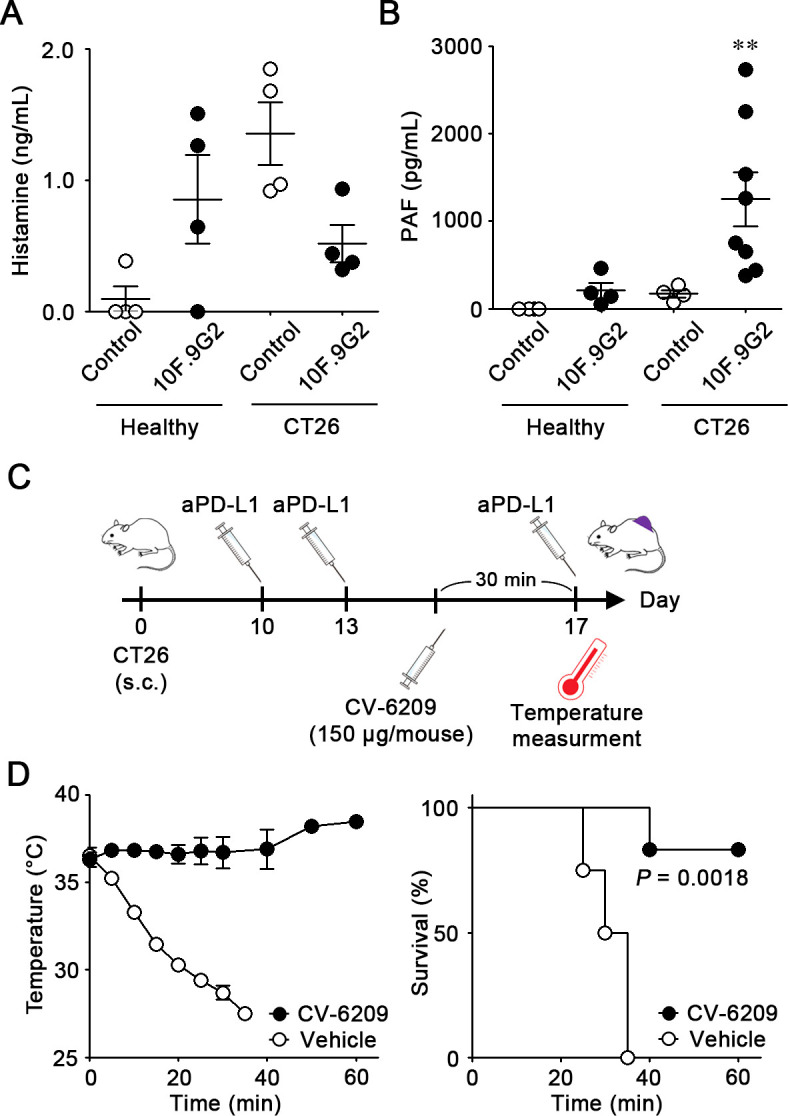
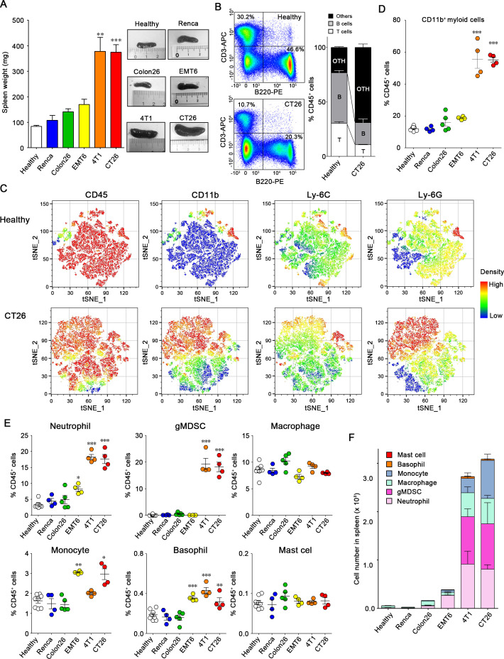
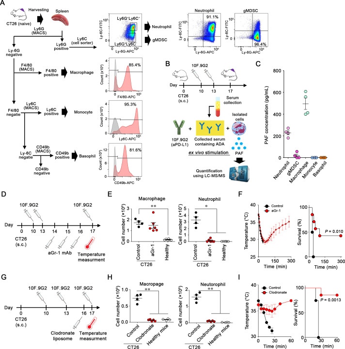
Methods: Healthy, murine colon 26, CT26, breast 4T1, EMT6, and renal RENCA tumor-bearing mice were treated with an anti-PD-L1 antibody (clone 10F.9G2). Symptoms of anaphylaxis were evaluated along with body temperature and mortality. The amounts of antidrug antibody and platelet-activating factor (PAF) in the blood were quantified via ELISA and liquid chromatography-mass spectrometry (LC-MS/MS). Immune cells were analyzed and isolated using a flow cytometer and magnetic-activated cell sorting, respectively.

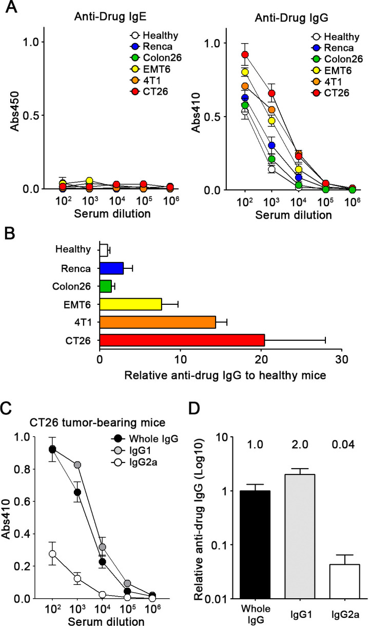
Results: Repeated administration of the anti-PD-L1 antibody 10F.9G2 to tumor-bearing mice caused fatal anaphylaxis, depending on the type of tumor model. After administration, antidrug immunoglobulin G (IgG), but not IgE antibodies, were produced, and PAF was released as a chemical mediator during anaphylaxis, indicating that anaphylaxis was caused by an IgG-dependent pathway. Anaphylaxis induced by 10F.9G2 was treated with a PAF receptor antagonist. We identified that neutrophils and macrophages were PAF-producing effector cells during anaphylaxis, and the tumor-bearing models with increased numbers of neutrophils and macrophages showed lethal anaphylaxis after treatment with 10F.9G2. Depletion of both neutrophils and macrophages using clodronate liposomes prevented anaphylaxis in tumor-bearing mice.

Conclusions: Thus, increased numbers of neutrophils and macrophages associated with cancer progression may be risk factors for anaphylaxis. These findings may provide useful insights into the mechanism of anaphylaxis following the administration of immune checkpoint inhibitors in human subjects.

Keywords: immunotherapy; translational medical research.


Conflict of interest statement

Competing interests: None declared.

Figures

Figure 1
Figure 1
Fatal anaphylaxis induced by sequential treatment with aPD-L1 mAb in CT26 tumor-bearing mice. (A) Experimental schedule for treatment of CT26 tumor-bearing mice with 10F.9G2. (B and C) Body temperature (B) and survival (C) of healthy (n=4), RENCA (n=5), Colon26 (n=4), EMT6 (n=5), 4T1 (n=7), and CT26 (n=11) tumor-bearing mice after the third injection of 10F.9G2. (D) Tumor volume of RENCA (n=5), Colon26 (n=4), EMT6 (n=5), 4T1 (n=4), and CT26 (n=6) tumor-bearing mice on day 17. (E) Experimental schedule for the treatment of CT26 tumor-bearing mice with 10F.9G2 and serum transfer. (F) body temperature and survival rate of CT26 tumor-bearing mice subjected to serum transfer were monitored after the first treatment with 10F.9G2 (n=6). Control mice (n=3) were treated with phosphate-buffered saline (PBS) instead of serum. The maximum temperature drop after treatment with 10F.9G2 is denoted as ΔT. Data are represented as mean±SE). N.S., not significant difference.
Figure 2
Figure 2
Exploring of subtype of antidrug antibodies to 10F.9G2. (A) Concentrations of the antidrug IgE and IgG against 10F.9G2 in the serum were evaluated using ELISA. healthy mice (n=4), Renca (n=4), EMT6 (n=4), 4T1 (n=9), and CT26 (n=6) tumor-bearing mice were treated with 10F.9G2 on days 10 and 13 postinoculation, and serum was collected on day 17. The x-axis represents the dilution of the serum sample, and the y-axis indicates the absorbance (Abs: A450 or A410). (B) Relative antidrug IgG levels in the serum. The serum dilution ratios resulting in Abs 410 of 0.5 when using antidrug IgG against 10F.9G2 were determined. (C) Evaluation of mouse IgG isotypes in antidrug antibodies using ELISA. CT26 (n=6) tumor-bearing mice were treated with 10F.9G2 on days 10 and 13 postinoculation and serum was collected on day 17. The concentrations of antidrug IgG, IgG1, and IgG2a against 10F.9G2 in the serum were evaluated using ELISA. The x-axis represents the dilution of the serum sample, and the y-axis indicates the absorbance (A410). (D) Relative antidrug IgG levels in the serum. Serum dilution ratios resulting in Abs 410 of 0.2 when using antidrug IgGs against 10F.9G2 were determined. Data are represented as mean±SE.
Figure 3
Figure 3
Evaluation of chemical mediators during anaphylaxis after 10F.9G2 administration. (A) Quantification of histamine in the serum by ELISA. Serum was collected 10 min after the third injection of 10F.9G2 from healthy (n=4) and CT26 tumor-bearing mice (n=4) treated with 10F.9G2 on days 10, 13, and 17 postinoculation. Serum from untreated mice was used as control (n=4). (B) Quantification of PAF in the serum by LC-MS/MS. Serum was collected as previously described from healthy (n=4) and CT26 tumor-bearing mice (n=8). **P<0.01. (C) Experimental schedule for the treatment of CT26 tumor-bearing mice with CV-6209 and 10F.9G2. CT26 tumor-bearing mice were treated with 10F.9G2 on days 10 and 13. CV-6209 (150 µg/mouse) was injected intraperitoneally 30 min before the third treatment with 10F.9G2. (D) Body temperature (left) and survival (right) of CT26 tumor-bearing mice treated with vehicle (n=4) or CV-6209 (n=6). Data are represented as mean±SE.
Figure 4
Figure 4
Increase in myeloid cells in the spleen of tumor-bearing mice. (A) Weight (left) and representative images (right) of the spleen from healthy (n=8), Renca (n=4), Colon26 (n=5), EMT6 (n=4), 4T1 (n=4), and CT26 (n=4) tumor-bearing mice on day 17 after inoculation. (B) Left: representative flow plots of CD45+ splenocytes from healthy and CT26 tumor-bearing mice stained with anti-CD3 and anti-B220 antibodies. Right: frequency of B cells (CD45+B220+), T cells (CD45+CD3+), and other fractions (CD45+B220CD3) in the spleens of healthy (n=4) and CT26 tumor-bearing mice (n=4). (C) Representative tSNE plots of splenocytes from healthy and CT26 tumor-bearing mice. tSNE analysis was performed using FlowJo software based on CD45, CD11b, Ly6C, and Ly6G expression. (D) Frequency of CD11b+ cells among CD45+ splenocytes in the spleens of healthy (n=8), Renca (n=4), Colon26 (n=5), EMT6 (n=4), 4T1 (n=4), and CT26 (n=4) tumor-bearing mice. (E) Frequency of neutrophils (CD11b+Ly6CintLy6G+), gMDSCs (CD11b+Ly6CLy6G+), macrophages (CD11b+Ly6GF4/80+), monocytes (CD11b+Ly6C+Ly6G), basophils (CD49b+FcεR1+), and mast cells (CD117+FcεR1+) in splenocytes. (F) Number of neutrophils, gMDSCs, macrophages, monocytes, basophils, and mast cells in the splenocytes. *P<0.05, **p<0.01, ***p<0.001, using one-way analysis of variance followed by Dunnett test (healthy mice vs tumor-bearing mice). Data are represented as mean±SE. gMDSCs, granulocytic myeloid-derived suppressor cells.
Figure 5
Figure 5
Neutrophils and macrophages cells release PAF during anaphylaxis. (A) Sorting of myeloid cells such as neutrophils, gMDSCs, macrophages, monocytes, and basophils from the spleen via magnetic-activated cell sorting (MACS) and a cell sorter. Histograms represent the purity of each subset. (B) Experimental schedule for ex vivo stimulation of myeloid cells isolated from the spleen of CT26 tumor-bearing mice. (C) The concentration of PAF released from isolated myeloid cells (n=4 per sample). (D) Experimental schedule for the treatment of CT26 tumor-bearing mice with 10F.9G2 on days 10, 13, and 17, and aGr-1 mAb on days 14, 15, and 16. (E) Cell numbers of macrophages and neutrophils in the spleen of healthy (n=6) and CT26 tumor-bearing mice 24 hours after treatment with either isotype control IgG (n=4) or aGr-1 mAb (n=6) on days 14, 15, and 16. (F) Body temperature (left) and survival (right) of CT26-tumor-bearing mice treated with either control IgG (n=4) or aGr-1 mAb (n=7) were monitored on day 17 after the third treatment with 10F.9G2. (G) Experimental schedule for the treatment of CT26 tumor-bearing mice with clodronate liposomes and 10F.9G2. (H) Cell numbers of macrophages and neutrophils in the spleen of healthy (n=4) and CT26 tumor-bearing mice 24 hours after treatment with either control (n=4) or clodronate (n=5) liposomes on day 16. (I) The body temperature (left) and survival (right) of CT26 tumor-bearing mice treated with either control (n=4) or clodronate (n=5) liposomes on day 16 were monitored after the third treatment with 10F.9G2 on day 17. Data represent mean±SE. *P<0.05, **p<0.01, ***p<0.001 by one-way analysis of variance followed by Bonferroni test. gMDSCs, granulocytic myeloid-derived suppressor cells; PAF, platelet-activating factor.

References

    1. Kemp SF, Lockey RF. Anaphylaxis: a review of causes and mechanisms. J Allergy Clin Immunol 2002;110:341–8. 10.1067/mai.2002.126811 - DOI - PubMed
    1. Finkelman FD, Khodoun MV, Strait R. Human IgE-independent systemic anaphylaxis. J Allergy Clin Immunol 2016;137:1674–80. 10.1016/j.jaci.2016.02.015 - DOI - PMC - PubMed
    1. Jiao D, Liu Y, Lu X, et al. . Macrophages are the dominant effector cells responsible for IgG-mediated passive systemic anaphylaxis challenged by natural protein antigen in BALB/c and C57BL/6 mice. Cell Immunol 2014;289:97–105. 10.1016/j.cellimm.2014.03.018 - DOI - PubMed
    1. Tsujimura Y, Obata K, Mukai K, et al. . Basophils play a pivotal role in immunoglobulin-G-mediated but not immunoglobulin-E-mediated systemic anaphylaxis. Immunity 2008;28:581–9. 10.1016/j.immuni.2008.02.008 - DOI - PubMed
    1. Murphy JT, Burey AP, Beebe AM, et al. . Anaphylaxis caused by repetitive doses of a GITR agonist monoclonal antibody in mice. Blood 2014;123:2172–80. 10.1182/blood-2013-12-544742 - DOI - PMC - PubMed

Publication types

Substances