. 2023 Apr;25(4):375-386.
doi: 10.1016/j.jcyt.2022.11.010. Epub 2022 Dec 20.

Soluble components from mesenchymal stromal cell processing exert anti-inflammatory effects and facilitate ischemic muscle regeneration

Affiliations

Soluble components from mesenchymal stromal cell processing exert anti-inflammatory effects and facilitate ischemic muscle regeneration

Wenbai Huang et al. Cytotherapy. 2023 Apr.

Abstract

Background aims: Skeletal muscle regeneration after severe damage is reliant on local stem cell proliferation and differentiation, processes that are tightly regulated by macrophages. Peripheral artery disease is a globally prevalent cardiovascular disease affecting millions of people. Progression of the disease leads to intermittent claudication, subsequent critical limb ischemia and muscle injury. Tissue-derived and ex vivo-expanded mesenchymal stromal cells (MSCs) for skeletal muscle regeneration have been studied, but pre-clinical and clinical results have not been consistent. As a result, the potential therapeutic efficacy and associated repair mechanisms of MSCs remain unclear. Numerous studies have demonstrated the vulnerability of delivered MSCs, with a precipitous drop in cell viability upon transplantation. This has prompted investigation into the therapeutic benefit of apoptotic cells, microvesicles, exosomes and soluble signals that are released upon cell death.

Methods: In this study, we characterized various components produced by MSCs after cell death induction under different conditions. We discovered anti-inflammatory and pro-regenerative effects produced by cell components following a freeze and thaw (F&T) process on macrophage polarization in vitro. We further investigated the underlying mechanisms of macrophage polarization by those components resulting from severe cell death induction.

Results: We found potent therapeutic effects from F&T-induced cell debris are dependent on the externalization of phosphatidylserine on the plasma membrane. In contrast, effects from the supernatant of F&T-induced cell death primarily depends on the released protein content. We then applied the F&T-induced cell supernatant to an animal model of peripheral artery disease to treat muscle injury caused by severe ischemia. Treatment with the F&T supernatant but not the vulnerable MSCs resulted in significantly improved recovery of muscle function, blood flow and morphology and inflammation resolution in the affected muscles 2 weeks after injury.

Conclusions: This study validates the therapeutic potential of F&T-induced supernatant obviating the need for a viable population from vulnerable MSCs to treat injury, thus providing a roadmap for cell-free therapeutic approaches for tissue regeneration.

Keywords: cell death; immunomodulation; macrophage polarization; mesenchymal stromal cells; muscle regeneration; stem cell therapy.


Conflict of interest statement

Declaration of Competing Interest The authors have no commercial, proprietary or financial interest in the products or companies described in this article.

Figures

Figure 1.
Figure 1.
(A) Schematic illustration of cell death induction in MSCs and the isolated cell components. (B) Images of the cell pellet with Annexin V and Propidium Iodide staining (Magnification, ×20; scale bar, 30 μm). (C) TEM images of F&T and heat supernatant confirming existence of EV-like vesicles in both cell components. (Scale bar: 100 nm).
Figure 2.
Figure 2.
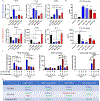
(A) Inflammation relevant gene expression of macrophages with different treatments and their expression relative to M0 macrophages (*p < 0.05). (B) VEGF and TNFα secretion from macrophages with different treatments under LPS stimulation (a, b, c, d, or e indicates the labeled group is significantly different from the group of a. M0 macrophages, b. LPS-M1 macrophages, c. F&T pellet, d. F&T supernatant, e. Heat pellet, respectfully; p < 0.05). (C) VEGF, TNFα and Nitrite production from macrophages with different treatments with or without IFNγ stimulation (*p < 0.05). n≥3 for all experiments. F: F&T; H: Heat; P: pellet; Sup.; S: supernatant. One-way ANOVA with Tukey’s multiple comparisons test was performed separately for IFNγ+ and IFNγ − conditions. (D) Summary table of the immunoregulatory effects of different cell components on macrophages under different conditions.
Figure 3.
Figure 3.
The protein secretion and gene expression of VEGF and TNFα from LPS-M1 macrophages in the presence or absence of anti-PtS antibody, # indicates the difference between the two group in the same condition and with or without the presence of anti-PtS antibody. Two-way ANOVA with Sidak’s multiple comparisons test was conducted to compare the means of the same treatment regarding the factor of PtS antibody presence, the control group without treatment and addition of PtS antibody was only presented as baseline reference and was not statistically analyzed. #p < 0.05, n = 3–4.
Figure 4.
Figure 4.
(A) Schematic illustration of F&T supernatant induced from different cell type. (B) TNFα secretion from LPS-M1 macrophages treated with F&T supernatant from different cell types at different doses. Two-way ANOVA with Sidak’s multiple comparisons test was conducted to compare the means at the same concentration across cell types (3T3 is NIH 3T3, a fibroblast cell line and RAW is RAW 264.7, a macrophage/monocyte cell line; * indicates difference between RAW sup. and MSC sup. and # indicates difference between RAW sup. and 3T3 sup.; n = 3–4). (C) TNFα secretion from LPS-M1 macrophages treated with F&T supernatant from MSCs after heating at different temperatures (n = 4). One-way ANOVA with Tukey’s multiple comparisons test was performed. (D) The relative concentration of the inflammation relevant proteins measured by protein array kit. Student’s t-test was performed for each protein (n = 4). *, # p < 0.05, **, ## p < 0.01, ***, ### p < 0.001, ****, #### p < 0.0001.
Figure 5.
Figure 5.
(A) Muscle tetanic force, (B) muscle mass, (C) muscle mass-normalized force of ischemic injured muscle after 14 days with different treatments (n = 5–8). (D) Representative histology images of H&E (top), Picrosirius Red (PSR; middle) and Oil Red O (ORO; bottom) staining of cross-sectioned muscle after ischemic injury (Scale bar: 100μm). (E) Quantitative analysis of percentage of myofibers with central nuclei (top) and average myofiber size (bottom) based on the H&E-stained sections (n=6). (F) Quantitative analysis of percentage of area of fibrosis (top) and neutral fat (bottom) based on the PSR and ORO-stained sections (n=6). One-way ANOVA with Tukey’s multiple comparisons test was performed. *p < 0.05, **p < 0.01, ***p<0.001, ****p<0.0001. (Intact indicates the intact muscles in contralateral hindlimb; Saline, MSCs, F&T sup. and Heat sup. are the ischemic muscles treated with saline, vulnerable MSCs, F&T supernatant and Heat supernatant)
Figure 6.
Figure 6.
(A) Relative blood flow with different treatments 14 days after the ischemic injury induction (n=5–8). (B) Representative images of laser speckle contrast imaging of relative blood flow of intact and affected foot. (C) Capillary density of cross-sectioned muscle with different treatments 14 days after ischemic injury based on the immunofluorescence staining of CD31 antibody (n=6). (D) Representative images of immunofluorescence staining (scale bar: 100μm). One-way ANOVA with Tukey’s multiple comparisons test was performed. *p < 0.05, ***
Figure 7.
Figure 7.
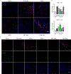
(A) Representative images of immunofluorescence imaging of CD68 (pink), CD206(green) and DAPI (blue) in muscle tissue. (B) Count of CD68+ and CD206+ cells (Scale bar: 50μm). One-way ANOVA with Tukey’s multiple comparisons test was performed. (a, b, c and d indicates the labeled group is significant different from the group of a. Intact, b. Untreated, c. Saline, d. Vulnerable MSCs; p < 0.05; n = 5–7). (C) Separated channels of CD68, CD206 and DAPI staining (Scale bar: 50μm).

References

    1. Kullo IJ, Rooke TW. CLINICAL PRACTICE. Peripheral Artery Disease. N Engl J Med 2016;374(9):861–871. doi:10.1056/NEJMcp1507631 - DOI - PubMed
    1. Virani SS, Alonso A, Aparicio HJ, et al. Heart Disease and Stroke Statistics-2021 Update: A Report From the American Heart Association. Circulation 2021;143(8):e254–e743. doi:10.1161/CIR.0000000000000950 - DOI - PubMed
    1. Tidball JG. Mechanisms of muscle injury, repair, and regeneration. Compr Physiol 2011;1(4):2029–2062. doi:10.1002/cphy.c100092 - DOI - PubMed
    1. Hammers DW, Rybalko V, Merscham-Banda M, Hsieh PL, Suggs LJ, Farrar RP. Anti-inflammatory macrophages improve skeletal muscle recovery from ischemia-reperfusion. J Appl Physiol (1985) 2015;118(8):1067–1074. doi:10.1152/japplphysiol.00313.2014 - DOI - PMC - PubMed
    1. Rybalko V, Hsieh PL, Ricles LM, Chung E, Farrar RP, Suggs LJ. Therapeutic potential of adipose-derived stem cells and macrophages for ischemic skeletal muscle repair. Regen Med 2017;12(2):153–167. doi:10.2217/rme-2016-0094 - DOI - PMC - PubMed

Publication types