. 2022 Dec 21;13(1):7850.
doi: 10.1038/s41467-022-35237-x.

Sourcing thermotolerant poly(ethylene terephthalate) hydrolase scaffolds from natural diversity

Affiliations

Sourcing thermotolerant poly(ethylene terephthalate) hydrolase scaffolds from natural diversity

Erika Erickson et al. Nat Commun. .

Abstract

Enzymatic deconstruction of poly(ethylene terephthalate) (PET) is under intense investigation, given the ability of hydrolase enzymes to depolymerize PET to its constituent monomers near the polymer glass transition temperature. To date, reported PET hydrolases have been sourced from a relatively narrow sequence space. Here, we identify additional PET-active biocatalysts from natural diversity by using bioinformatics and machine learning to mine 74 putative thermotolerant PET hydrolases. We successfully express, purify, and assay 51 enzymes from seven distinct phylogenetic groups; observing PET hydrolysis activity on amorphous PET film from 37 enzymes in reactions spanning pH from 4.5-9.0 and temperatures from 30-70 °C. We conduct PET hydrolysis time-course reactions with the best-performing enzymes, where we observe differences in substrate selectivity as function of PET morphology. We employed X-ray crystallography and AlphaFold to examine the enzyme architectures of all 74 candidates, revealing protein folds and accessory domains not previously associated with PET deconstruction. Overall, this study expands the number and diversity of thermotolerant scaffolds for enzymatic PET deconstruction.


Conflict of interest statement

U.S. Patent Application No.: 63/297,529 was submitted by the National Renewable Energy Laboratory (NREL), operated by Alliance for Sustainable Energy, LLC. The inventors include E.E., J.E.G., C.M.P., G.T.B., and J.E.M. The other authors declare no competing interests.

Figures
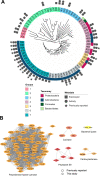
Fig. 1
Fig. 1. Bioinformatics and machine learning to derive PET hydrolase sequences from natural diversity.
A PET hydrolase candidates (74 total) selected by HMM and ML shown with a minimum-evolution phylogenetic tree. Sequences retrieved from environmental (meta)genomes in JGI IMG with lower HMM scores (groups 1–3) are notably diverse compared to the sequences that comprise the rest of the tree (groups 4–7). The symbols around the tree show expression, activity, and previously reported PET activity. Full organism names and accession numbers are shown in Supplementary Table 9, and sequence identity between these 74 sequences and previously reported PETases is shown in Supplementary Table 8. A maximum-likelihood phylogenetic tree of all experimentally confirmed PET hydrolases is shown in Supplementary Fig. 1. B Sequence Similarity Network (SSN) of PET hydrolases with experimentally confirmed PET hydrolase activity, including sequences examined in this study and previously reported PETases. Edges represent pairwise BLAST similarity with E-value < 1e–10. The SSN clusters are consistent with the associated families in the ESTHER database, and show that most reported PET hydrolases fall in the polyester-lipase-cutinase family. We note that these clusters are different from phylogenetic groups in (A). Full details of experimentally verified PET hydrolases are shown in Supplementary Tables 1 and 10.
Fig. 2
Fig. 2. Enzyme activities.
Heat map profiles of pH and temperature screening for hydrolytic activity on amorphous PET film by a diverse selection of 19 candidate enzymes and a positive control enzyme, LCC ICCG. The heat map gradient indicates the extent of measured product release up to 500 mg/L of total aromatic products after 96 h reaction time, and is reported as the average of reactions performed in triplicate (n = 3). Each heat map displays the reaction conditions utilized (citrate at pH 6.0, NaH2PO4 at pH 7.0, NaH2PO4 at pH 7.5, HEPES (H) at pH 7.5, bicine at pH 8.0, and glycine at pH 9.0), and reaction temperature (30, 40, 50, 60, or 70 °C). The heat maps for all other enzymes tested on amorphous PET film are shown in Supplementary Fig. 3. Source data are provided as a Source Data file.
Fig. 3
Fig. 3. Substrate selectivity varies across PET morphologies.
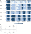
A Heat map profiles of pH and temperature screening for hydrolytic activity on 3 PET substrate morphologies, the same amorphous PET film presented in Fig. 2, as well as an amorphous PET powder and a crystalline PET powder, using a subset of 9 candidate enzymes and positive control enzyme, LCC ICCG. The heat map gradient indicates extent of measured product release up to 500 mg/L of total aromatic products after 96 h reaction time, and is reported as the average of reactions performed in triplicate (n = 3). Each heat map displays the reaction conditions utilized (citrate at pH 6.0, NaH2PO4 at pH 7.0, NaH2PO4 at pH 7.5, HEPES (H) at pH 7.5, bicine at pH 8.0, and glycine at pH 9.0), and reaction temperature (30, 40, 50, 60, or 70 °C). The heat maps for all other enzymes tested on the 3 PET substrate morphologies are shown in Supplementary Fig. 6. Source data are provided the a Source Data file. B Log-plot of the sum of aromatic products measured after 168 h reaction time using amorphous PET film (aFilm, black squares), crystalline PET powder (cryPow, open circles) and amorphous PET powder (aPow, gray circles) as substrates. Reaction conditions used for time course experiments correspond to the pH and temperature resulting in the highest product release observed in amorphous PET film screening reactions, which are listed in Supplementary Table 13. Ratios of product release observed from hydrolysis reactions for each PET substrate morphology pairwise comparison, demonstrating differences in substrate selectivity for each selected enzyme is presented in Supplementary Fig. 9. For all enzymatic reactions shown in A, B, the enzyme loading was 0.7 mg enzyme/g PET and the solids loading was 2.9% (29  g/L). The reaction products were quantified with UHPLC, and the results show the sum of aromatic products, including BHET, MHET, and TPA. All reactions were conducted in triplicate (n = 3). Error bars represent standard deviation and are centered on the average of the three reaction measurements. Source data are provided as a Source Data file.
Fig. 4
Fig. 4. Structural diversity of PET-active and representative enzymes from phylogenetic groups.
All structural models are shown to scale, rendered as cartoons with transparent accessible surface areas and putative active sites highlighted with the Ser-His-Asp catalytic triad in red sticks. A PET hydrolase scaffolds identified from mesophilic (top, I. sakaiensis PETase, PDB ID 6EQE) and thermophilic (middle, LCC, PDB ID 4EB0, and bottom, T. fusca cutinase 1 DSM44342 (703; PDB ID 7QJR)) sources occupy a narrow structural space with highly conserved α/β hydrolase folds. B A selection of representatives from more distant phylogenetic groups reveals multiple additional and alternative structural features with substantial increases (102) and reductions (307) in the core fold. C Several additional distinct domains were revealed, including a Peripheral Subunit-Binding Domain (PSBD) and a Family 35 carbohydrate binding module (CBM).
Fig. 5
Fig. 5. Increasing degrees of structural diversity across phylogenetic groups.
A Conserved canonical folds with surface residue changes in groups 5 and 6. Electrostatic surface representations are colored with a gradient from red (acidic) at −7 kT/e to blue (basic) at 7 kT/e (where k is Boltzmann’s constant, T is temperature, and e is the charge on an electron). The general location of active site cleft is indicated with a star. Known (LCC) and predicted catalytic triad residues are shown as stick representations in the corresponding images below. B Accessory lid domains in group 2 enzymes. Examples of alternative lid domains are highlighted in green. C Mini-PETases are created from large core deletions to the canonical fold. LCC is shown in the middle column (yellow) as a cartoon with the catalytic triad highlighted in red, and a surface representation below with a PET trimer (blue) docked in the active site cleft. A comparison with 307 on the left (cartoon above shown without the lid domain for clarity) reveals the extent of the core deletion, removing four of the eight β-strands and corresponding helices. A comparison with 305 on the right reveals an almost complementary set of deletions. These major rearrangements generate alternative binding clefts and docking studies predict vastly different binding modes (PET trimers in blue). Superpositions of the three enzymes in this panel are depicted in Supplementary Fig. 19. D An alternative enzyme family for PET hydrolysis. The enzymes 101 (left) and 102 (right) are colored according to the 3-domain arrangement in the Geobacillus stearothermophilus carboxylesterase EST55 (PDB ID 2OGT). Both enzymes display a truncated version of the catalytic domain (pink) compared to EST55 (Supplementary Fig. 20) and have modified versions of the α/β domain (blue). Only enzyme 101 has a version of the regulatory domain, the absence of which in 102 disrupts the formation of the canonical active site (locations highlighted with red dashes). While the catalytic Ser and Glu residues are conserved between EST55 and 101 (pink and yellow sticks), there is no direct substitute for the His residue. In enzyme 102, only the catalytic Ser is position is conserved (Supplementary Fig. 20).

References

    1. Sinha V, Patel MR, Patel JV. PET waste management by chemical recycling: a review. J. Polym. Environ. 2010;18:8–25. doi: 10.1007/s10924-008-0106-7. - DOI
    1. Rahimi A, García JM. Chemical recycling of waste plastics for new materials production. Nat. Rev. Chem. 2017;1:1–11. doi: 10.1038/s41570-017-0046. - DOI
    1. Ellis LD, et al. Chemical and biological catalysis for plastics recycling and upcycling. Nat. Catal. 2021;4:539–556. doi: 10.1038/s41929-021-00648-4. - DOI
    1. Martín, A. J., Mondelli, C., Jaydev, S. D. & Pérez-Ramírez, J. Catalytic processing of plastic waste on the rise. Chem 7, 1487–1533 (2021).
    1. Wei R, Zimmermann W. Biocatalysis as a green route for recycling the recalcitrant plastic polyethylene terephthalate. Micro. Biotechnol. 2017;10:1302–1307. doi: 10.1111/1751-7915.12714. - DOI - PMC - PubMed

Publication types