. 2022 Dec 21;13(1):7848.
doi: 10.1038/s41467-022-34197-6.

Quantitative phase contrast imaging with a nonlocal angle-selective metasurface

Affiliations

Quantitative phase contrast imaging with a nonlocal angle-selective metasurface

Anqi Ji et al. Nat Commun. .

Abstract

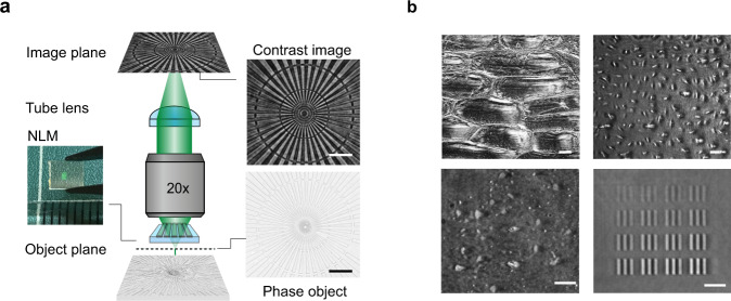
Phase contrast microscopy has played a central role in the development of modern biology, geology, and nanotechnology. It can visualize the structure of translucent objects that remains hidden in regular optical microscopes. The optical layout of a phase contrast microscope is based on a 4 f image processing setup and has essentially remained unchanged since its invention by Zernike in the early 1930s. Here, we propose a conceptually new approach to phase contrast imaging that harnesses the non-local optical response of a guided-mode-resonator metasurface. We highlight its benefits and demonstrate the imaging of various phase objects, including biological cells, polymeric nanostructures, and transparent metasurfaces. Our results showcase that the addition of this non-local metasurface to a conventional microscope enables quantitative phase contrast imaging with a 0.02π phase accuracy. At a high level, this work adds to the growing body of research aimed at the use of metasurfaces for analog optical computing.


Conflict of interest statement

A.J., J.S. and M.B. have submitted a patent application (PCT/US2020/061616) based on the ideas presented in this work. There are no other competing interests.

Figures

Fig. 1
Fig. 1. A nonlocal metasurface (NLM) patterned on a microscope slide enables phase contrast imaging.
a A schematic showing how a classic bright field microscope can be transformed into a phase imaging setup simply by inserting an NLM on top of the phase object. The flat optical component, a commercial phase object and the corresponding phase contrast image are shown next to the microscope schematic. b NLM phase contrast images of onion cells (top left), human osteosarcoma cells (U-2 OS, top right), a dense array of exfoliated transparent hexagonal boron nitride (hBN) flakes (bottom left), and arrays of metasurface elements comprised of silicon nitride pillars with different diameters (bottom right). All scale bars in a and b correspond to 25 µm length.
Fig. 2
Fig. 2. Optical image processing can be achieved with a NLM that serves as a designer angular filter.
a Top: Our proposed phase contrast system utilizing an NLM-based angular filter. Bottom: A schematic of a conventional 4 f image processing system with a Fourier filter is shown for reference. b A cross sectional schematic of the NLM. Incident transverse magnetic (TM) polarized light can take a direct (1) and indirect, resonant pathway (2) through this optical element. The corresponding field distributions are shown underneath the schematic. c The simulated transmittance spectrum of our NLM at normal incidence shows a pronounced dip that results from destructive interference between the direct and indirect pathways on resonance. d Simulated angular transmittance spectra of the NLM on and near the resonant wavelength λ0. The case without the surface relief grating is shown for reference. e Simulated phase shift produced by the NLM for incident light at different angles. Insets show positive contrast images (blue box) and negative contrast images (green box) that were taken below and above the resonant wavelength, respectively.
Fig. 3
Fig. 3. Experimental validation of the angular response of our NLM.
a Experimental map of the metasurface transmittance versus wavelength and incident angle that reflects the dispersion of the transverse magnetic (TM) mode of the single-mode silicon nitride slab corrugated with the grating. b Simulated map of the transmittance that closely matches the experiments in panel a. c Angle-dependent transmittance of the NLM at three wavelengths near the resonant wavelength λ0 = 630 nm. (dashed line: simulation, solid line: experiment) d Normal incidence transmission images of the NLM taken at various illumination wavelengths around λ0 = 630 nm show the reduced transmission across the entire patterned area on resonance (NKT supercontinuum source, TM polarized). e Reflection optical microscope image of the NLM.
Fig. 4
Fig. 4. A quantitative comparison of conventional and NLM phase contrast images.
a Bright field image of a phase-contrast calibration sample. b Phase contrast image of the calibration sample recorded with Zernike’s method. c, d. c Simulated, and d. experimental phase contrast images taken with the NLM. e Line profiles of the bars in group 7 of the calibration sample (indicated by the red line in panel a) allow for a quantitative comparison of the different imaging approaches. From top to bottom, the profiles are extracted from panels a, b, c, and d, respectively. C represents the contrast values. All images are measured on a polymer USAF target (n = 1.5) that has 200 nm thickness.
Fig. 5
Fig. 5. Quantitative phase retrieval using an NLM.
a Transmission and phase delay from 2D periodic arrays of Si3N4 nanorods with heights of h = 230 nm and lattice constant a = 360 nm. The inset shows the structure of a single cell. The error bars show a reference of ±0.02π phase difference with respect to the measured phase. b SEM image of different area of the fabricated metasurface. c Simulated NLM phase contrast image. d Measured NLM phase contrast image. Scale bar corresponds to 20 µm.

References

    1. LeCun Y, Bengio Y, Hinton G. Deep learning. Nature. 2015;521:436–444. doi: 10.1038/nature14539. - DOI - PubMed
    1. Ren S, He K, Girshick R, Sun J. Faster R-CNN: Towards Real-Time Object Detection with Region Proposal Networks. IEEE Trans. Pattern Anal. Mach. Intell. 2017;39:1137–1149. doi: 10.1109/TPAMI.2016.2577031. - DOI - PubMed
    1. Russakovsky O, et al. ImageNet Large Scale Visual Recognition Challenge. Int. J. Computer Vis. 2015;115:211–252. doi: 10.1007/s11263-015-0816-y. - DOI
    1. Wetzstein G, et al. Inference in artificial intelligence with deep optics and photonics. Nature. 2020;588:39–47. doi: 10.1038/s41586-020-2973-6. - DOI - PubMed
    1. Guo Q, et al. Compact single-shot metalens depth sensors inspired by eyes of jumping spiders. Proc. Natl Acad. Sci. 2019;116:22959–22965. doi: 10.1073/pnas.1912154116. - DOI - PMC - PubMed

Publication types